


 7 / 36

Page 75
conferenceseries
.com
Volume 4
Toxicology: Open Access
ISSN: 2476-2067
Toxicology Congress 2018
March 12-14, 2018
March 12-14, 2018 Singapore
14
th
World Congress on
Toxicology and Pharmacology
Evaluation of oxidative stress effect for
in vitro
maturated cow oocytes through gene expression quantification
Camelia Tulcan, Calin Mircu, Ioan Hutu, Cornel Balta, Popescu Sorina, Marc Simona, Alexa Ersilia and Oana-Maria Boldura
Banat’s University of Agricultural Sciences and Veterinary Medicine Timisoara, Romania
I
n the current assisted reproductive practice in cows the IVF technique is used in
increasing proportion. However, the used methods are not always standardized
and are needed to be improved. The first challenge in optimization of IVF techniques
is obtaining mature oocytes by growing them in culture media and by this to preserve
their high fertilization quality. A crucial factor in improving IVF results is the
prevention of oocytes from
in vitro
cultivation stress effects. In the presented study
the beneficial effect of antioxidant supplementation in maturation culture media of
cow oocytes was evaluated by apoptotic genes expression quantification. The oocytes
were cultivated for 24 hours on conventional (control variant), supplemented with
rosmarinic acid (RA variant) and ascorbic acid (C variant), maturation media.
The oocytes were classified in three quality classes by morphological observation
from which the total RNA was isolated. Quantitative PCR technique was used for
quantification of BAX and BCL2 apoptotic genes expression. Results of qPCR were
interpreted by Δ (ΔCt) method. The ratio BCL2/BAX was considered as an indicator
of maturated oocytes homeostasis. Antioxidants culture media supplementation
resulted in a better expansion of cumulus cells. The level of expression of the BAX
gene has an increasing trend in all COC’s, inversely proportional to oocyte quality,
indicating the overcoming of cell adaptation process for the inferior class. Regarding
the BCL2 gene, significantly higher expression levels can be observed in class
I oocytes supplemented with antioxidants. The level of maintenance of cell homeostasis, as reflected by the ratio of BAX/
BCL-2, with a value above 7, indicates that apoptotic processes have been installed in all class III oocytes. Supplementation
with antioxidants exerts a beneficial effect on inferior class cells, which have a high stress level, to some extent assuring their
protection, indicating the effectiveness of administering this supplement.
References
1. Mbemya G T (2017) Reports on
in vivo
and
in vitro
contribution of medicinal plants to improve the female reproductive
function.
Reprod Clim.
; 32(2): 109-119.
2. Roychoudhury S, Agarwal A, Virk G, Cho C-L (2017) Potential role of green tea catechins in the management of oxidative
stress-associated infertility. Reproductive BioMedicine Online; 34(5): 487-498.
3. Kang J T, Moon J H, Choi J I, Park S J, Kim S J, Saadeldin I M, Lee B C (2016) Effect of Antioxidant Flavonoids (Quercetin
and Taxifolin) on
In vitro
Maturation of Porcine Oocytes, Asian Australias.
J. Anim. Sci.
; 29(3): 352-358.
4. Luno V, Gil L, Olaciregui M, Gonzalez N, Jerez R A, Blas I (2014) Rosmarinic acid improves function and
in vitro
fertilising
ability of boar sperm after cryopreservation,
Cryobiology
; 69(1): 157-162.
5. Sadeesh E, Shah F, Balhara A K, Thirumaran S MK, Yadav S, Yadav P S (2014) Effect of growth factor and antioxidant on
in
vitro
maturation of oocytes and cleavage rates of
in vitro
produced Indian buffalo
(Bubalus bubalis)
embryos.
Veterinarski
Arhiv;
84(5): 459-474.
Biography
Camelia Tulcan is an Assistant Professor at Biochemistry Department, Faculty of Veterinary Medicine, Timisoara and Coordinator of Antioxidant Research Lab-
Horia Cernescu Research Unit. She has expertise in oxidative stress evaluation in different physiological or pathological condition and was involved in management
team of research infrastructure project and in implementation of quality management systems.
cameliatulcan@usab-tm.ro, tulcancamelia@gmail.comCamelia Tulcan et al., Toxicol Open Access 2018, Volume 4
DOI: 10.4172/2476-2067-C1-006
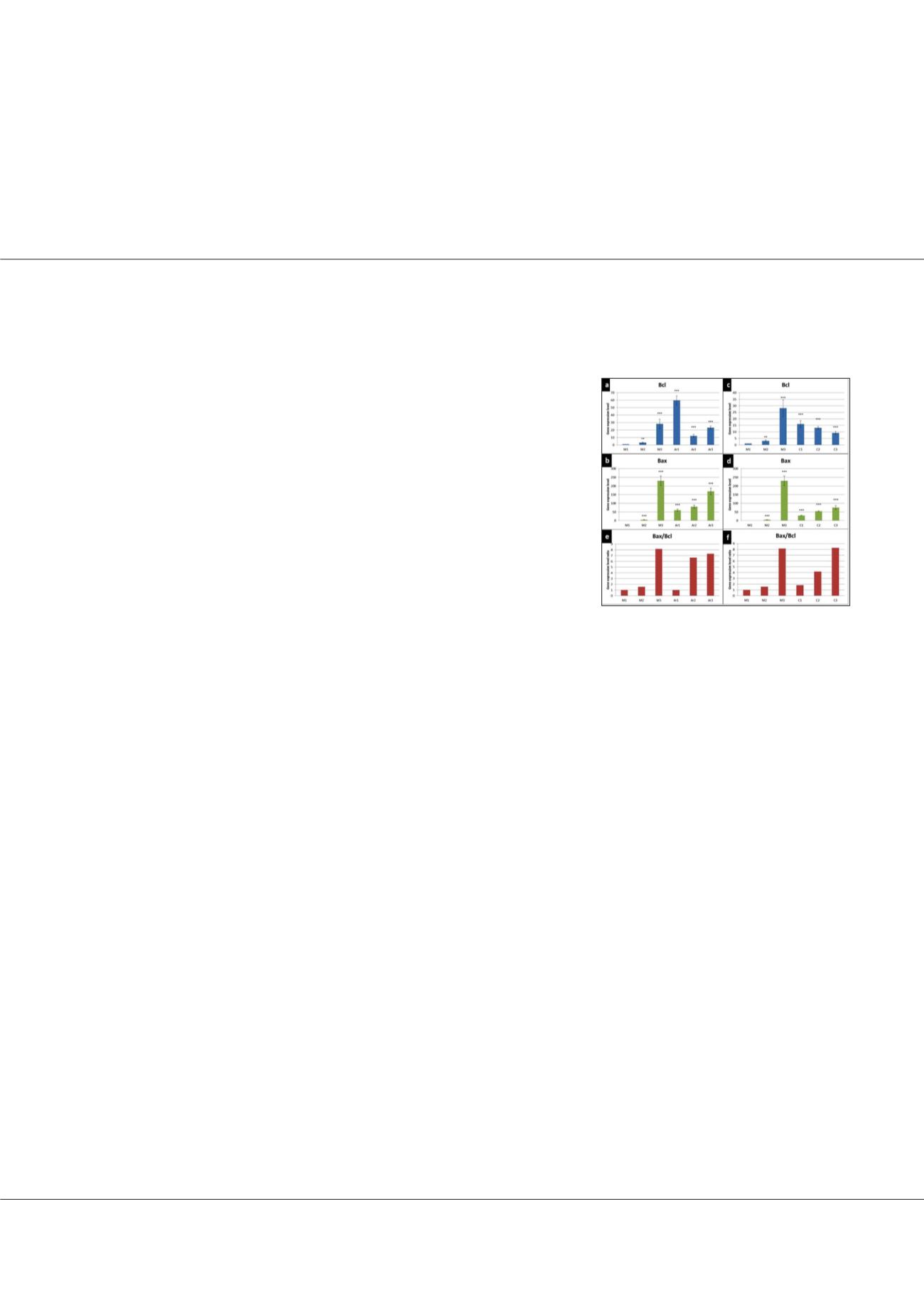
Figure-1:
BAX and BCL2 gene expression
maturated oocytes. a- BCL2 gene expression for AR
samples; b - BAX gene expression for AR samples;
c - BCL2 gene expression for C samples; b - BAX
gene expression for C samples; e - BAX/BCL2 genes
expression ratio for AR samples; d - BAX/BCL2
genes expression ratio for C samples.