


 84 / 103

Page 123
Notes:
conferenceseries
.com
Volume 10, Issue 8 (Suppl)
J Proteomics Bioinform, an open access journal
ISSN: 0974-276X
Structural Biology 2017
September 18-20, 2017
9
th
International Conference on
Structural Biology
September 18-20, 2017 Zurich, Switzerland
Ozge Sensoy, J Proteomics Bioinform 2017, 10:8(Suppl)
DOI: 10.4172/0974-276X-C1-0100
Understanding differential selectivity of arrestins toward the phosphorylation state of G-protein-
coupled receptors
Ozge Sensoy
Istanbul Medipol University, Turkey
A
rrestins (Arrs) are a family of four proteins (Arr1- 4) which mediate G-protein-coupled receptor (GPCR) desensitization
and internalization by coupling to active and phosphorylated receptor. Recently, they have also been shown to mediate
GPCR-independent signaling pathways. The specific functions of Arrs (desensitization vs. G-protein-independent signaling)
can be regulated by differential phosphorylation of the receptor, which is known as the
phosphorylation barcode
. The molecular
mechanism responsible for formation of a high-affinity complex between an Arr subtype and a GPCR having a certain
phosphorylation pattern remains elusive but is crucial for directing the subtype towards a specific functional role, and hence
paves the way for development of safer therapeutics with fewer side-effects. As a first step in that direction, we have started
with elucidating the activation mechanism of Arr subtypes by carrying out comparative molecular dynamics (MD) studies
of the two members of the family, namely Arr1 and Arr3, which exhibit the largest differences in terms of phosphorylation
selectivity. In addition, we also modeled and simulated Arr1-R175E mutant, which is known to be constitutively active, and
compared it to Arr1 and Arr3 to detect activation-related rearrangements. We found novel structural elements that had not
been considered before as determinants for activation and can be targeted with drugs for functional modulation. The emerging
model also proposes that activation of Arr1-R175E is connected to perturbation of the well-known region, namely, the polar-
core, whereas no changes were observed in that region in Arr3 despite the presence of other activation-related changes. With
that, we could propose a structural model to explain the molecular mechanism responsible for markedly reduced selectivity of
Arr3 towards phosphorylated GPCRs. Finally, knowledge achieved in this study can also be utilized to modulate Arr binding
to GPCRs under disease conditions such as otozomal dominant disorders and congestive heart failure.
Biography
Ozge Sensoy being a Computational Biophysicist, her research has focused on understanding molecular mechanisms of biologically important problems and
providing mechanistic insight at the molecular level. In particular, she has been working with GPCRs and their interacting partners which are responsible for cellular
signaling. She works in close collaboration with medicinal chemists to direct them for effective molecular designs. In addition, she is also responsible for testing the
efficacy of these molecules
in silico
before transferring them to either
in vitro
or
in vivo
studies. Recently, she has been awarded an international COST (European
Cooperation in Science and Technology) grant which is based on developing heterobivalent molecules capable of binding more than one target for treatment of
symptoms of Parkinson’s disease.
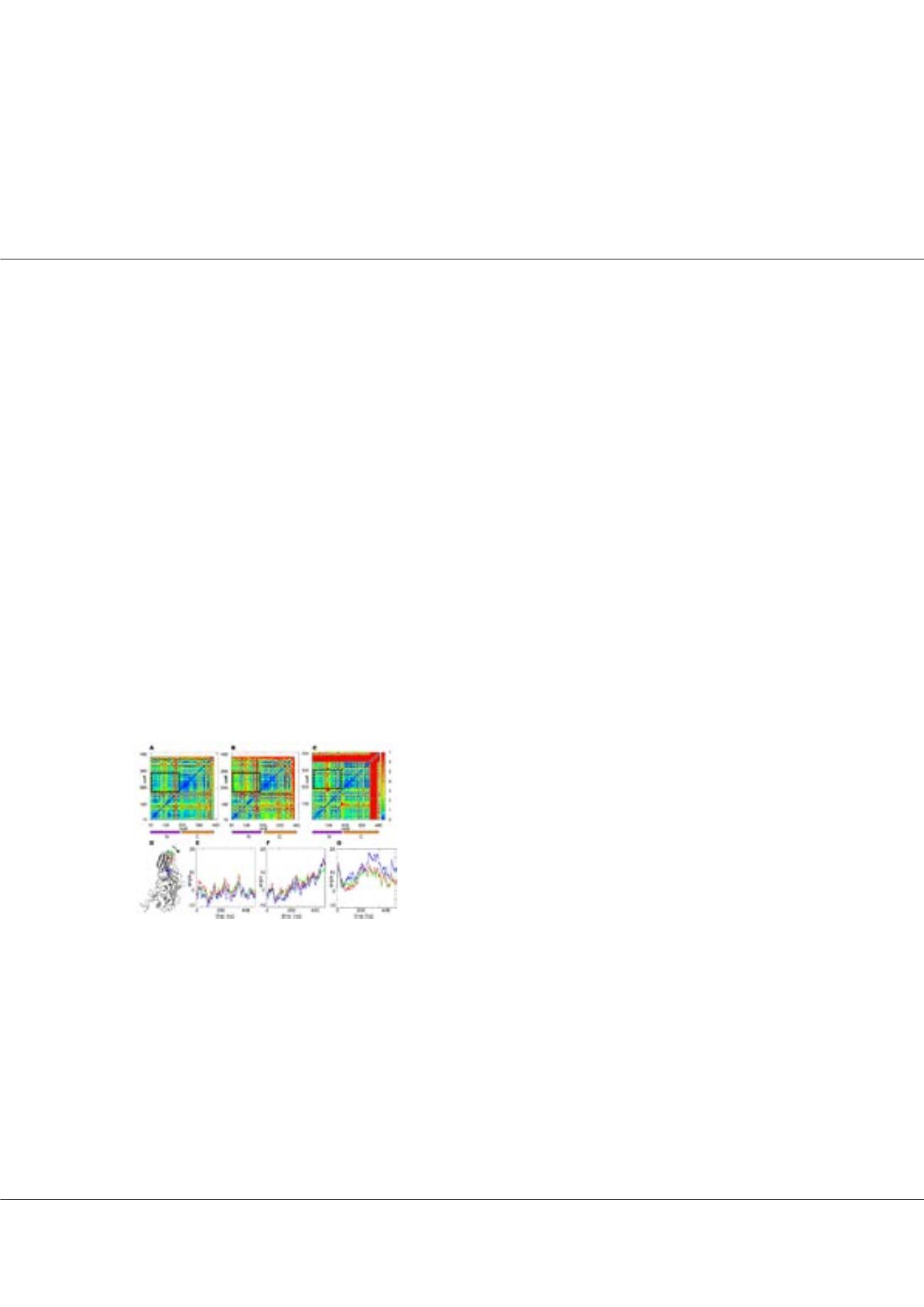
osensoy@medipol.edu.trFigure1:
Top, Distance fluctuation maps calculated for Cα-Cα distances along the
MD trajectories for (A) Arr1, (B) Arr1-R175E (B) and (C) Arr3. N and C-domains
are identified by stripes. The dashed rectangles highlight the distance fluctuations
in the N-C interface region. Bottom, Panel D: View of Arr3, with rotation axis
perpendicular to the Picture plane and passing through Cα of I321, depicted in
black (I306 in Arr1); rotation on this axis in the direction of the arrow corresponds
to an increase in the rotation angle. Cα atoms used in quantifying the rotation
along the MD trajectory are represented in blue, red, and green. Panels (E-G):
Time dependent rotation of the selected residues in Arr1-WT (E), in Arr1R175E
(F) and Arr3(G).