


 19 / 103

Page 51
conferenceseries
.com
Volume 10, Issue 8 (Suppl)
J Proteomics Bioinform, an open access journal
ISSN: 0974-276X
Structural Biology 2017
September 18-20, 2017
9
th
International Conference on
Structural Biology
September 18-20, 2017 Zurich, Switzerland
Cryo-electron microscopy grid preparation from nanoliter-sized protein samples and single-cell
extracts
Thomas Braun, Stefan Arnold, Stefan Albiez, Andrej Bieri, Claudio Schmidli, Anastasia Syntychaki, Luca Rima, Nadia Opara, Shirley Müller, Kenneth N
Goldie, Mohamed Chami
and
Henning Stahlberg
University of Basel, Switzerland
C
ryo-electron microscopy (cryo-EM) sample preparation techniques ensure that biological specimens can be investigated
at physiological conditions in the electron microscope. However, these preparation methods suffer from extensive blotting
steps leading to a massive loss of sample and sometimes to partial denaturation of sensitive protein complexes. We have
developed a simple method for the almost lossless conditioning and preparation of nanolitre-volumes of biological samples for
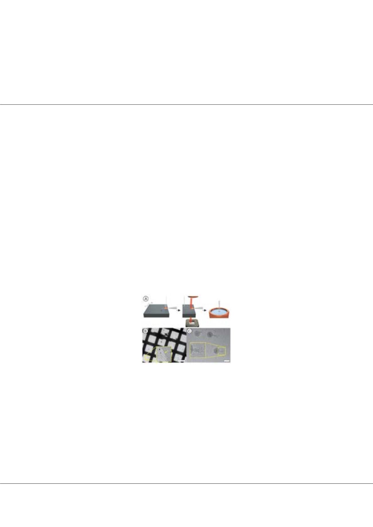
EM. The method does not involve any blotting steps. Amicrocapillary is used to aspirate 3 to 20 nanoliters of sample, depending
on the experiment. In the figure, the sample is applied (left) and spread (center) on the EM-grid. Real-time monitoring allows
the thickness of the water film to be assessed and decreased to the optimum value prior to vitrification (right). We prepared
cryo-EM grids of various samples, e.g., bacteriophages and soluble proteins as shown in Figure 1B and C, to demonstrate
the usefulness and general applicability of the method. We also showed that high-resolution 3D structures can be calculated
from single-particle preparations of a soluble protein. In addition to cryo-EM grid preparation, the versatile method allows
nanoliter-sized sample volumes to be conditioned for EM, e.g., negatively stained with heavy metal salts or embedded in
trehalose. In addition, we combine the new sample preparation method with a single cell lysis device for adherent eukaryotic
cells and image the aspirated cell contents by TEM. To demonstrate the usefulness of this new visual proteomics approach we
visualized the changes occurring in single cell proteomes upon heat shocking the cells. Furthermore, we have developed a
protein-fishing method based on a magnetic trap and photo-cleavable composite material, to ‘fish’ untagged proteins from cell
lysate by antibodies. This allows target proteins to be isolated from approx. 40,000 cells in 90 min and analyzed by EM.
Biography
Thomas Braun has received his PhD in 2002 in Biophysics from the Biozentrum, University of Basel, Switzerland. During his PhD thesis, he has applied high-
resolution electron microscopy and digital image processing to study the structure and function of membrane proteins. Subsequently, he has worked on nano-
mechanical sensors to characterize the mechanics of membrane proteins at the Institute of Physics, University Basel and the CRANN, Trinity College Dublin,
Ireland. He has been working at the Center for Cellular Imaging an Nano Analytics (Biozentrum, University of Basel, Switzerland) since 2009 and is developing new
methods for electron microscopy, single cell analysis and nano-mechanical sensors for biological applications.
thomas.braun@unibas.chThomas Braun et al., J Proteomics Bioinform 2017, 10:8(Suppl)
DOI: 10.4172/0974-276X-C1-0100
Figure1:
(A) Cryo-EM grid preparation from nanoliter-sized samples (see main text). (B) Example overview, yellow arrows
indicate borders of the vitreous ice. Scale bar: 100 µm. (C) Test-sample containing apoferritin particles and bacteriophages at high
magnification and defocus (to increase contrast). Inset: twofold enlargement of the indicated region. Scale bar: 80 nm.