


 15 / 103

Page 47
conferenceseries
.com
Volume 10, Issue 8 (Suppl)
J Proteomics Bioinform, an open access journal
ISSN: 0974-276X
Structural Biology 2017
September 18-20, 2017
9
th
International Conference on
Structural Biology
September 18-20, 2017 Zurich, Switzerland
The native (Sulfur) SAD method in photon factory
Toshiya Senda
High Energy Accelerator Research Organization, Japan
C
rystallography has been a major method to determine 3D structures of biological macromolecules at atomic resolution.
While a newmethod with cryo-EM is becoming another major technique for the 3D structure analysis, crystallography still
has some advantages. Recently, many crystal structures of biological macromolecules are determined by MAD/SAD method
with seleno-methionine proteins (SeMet-proteins). Since selenium has an X-ray absorption edge near 1Å, it is convenient to
utilize in the MAD/SAD method. While this technique is useful, crystallographers need to prepare SeMet-proteins. If we can
develop a method to solve the phase problem without using SeMet-proteins, it would be highly useful for crystallographers. So,
we have tried to develop the native SAD (or sulfur SAD) method, in which anomalous signals from sulfur atoms in the native
protein are utilized. However, there are some problems in the native SAD method. First, sulfur gives only weak anomalous
signals with X-ray typically utilized in protein crystallography (X-ray wavelength of around 1Å). To increase the anomalous
signals, we need to use a longer wavelength X-ray than usual. However, since a longer wavelength X-ray shows significant
absorption by air, solvent, protein etc., data quality is degraded by the absorption. The native SAD method, therefore, requires
a specific system for high quality data collection. To achieve routine utilization of the native SAD method, we have developed
a beamline (BL-1A) dedicated for the native SAD method. In BL-1A, we can utilize a long wavelength X-ray (1.9-3.5 Å).
Furthermore, the goniometer and X-ray detector are installed inside a He chamber to prevent the absorption problem. Our
system enables us to solve crystal structures of proteins by the native SAD method. In the presentation, we will present several
examples of crystal structure determination with native SAD. Also, we will mention our unique method for crystal freezing to
collect high quality diffraction data required in native SAD experiments.
Biography
Toshiya Senda has completed his PhD from Nagaoka University of Technology (Niigata, Japan) in 1995. He was a Research Associate in Nagaoka University of
Technology (1995-2001) and a Senior Researcher in Institute of Advanced Industrial Science and Technology (2001-2012). Now, he is the Director/Professor of
Structural Biology Research Center of High Energy Accelerator Research Organization (KEKI) in Japan. He was awarded the CrSJ (Crystallographic society of
Japan) award in 2014 (Structural biology studies of CagA from
Helicobacter pylori
and histone chaperon CIA/ASF1).
toshiya.sen
da@kek.jpToshiya Senda, J Proteomics Bioinform 2017, 10:8(Suppl)
DOI: 10.4172/0974-276X-C1-0100
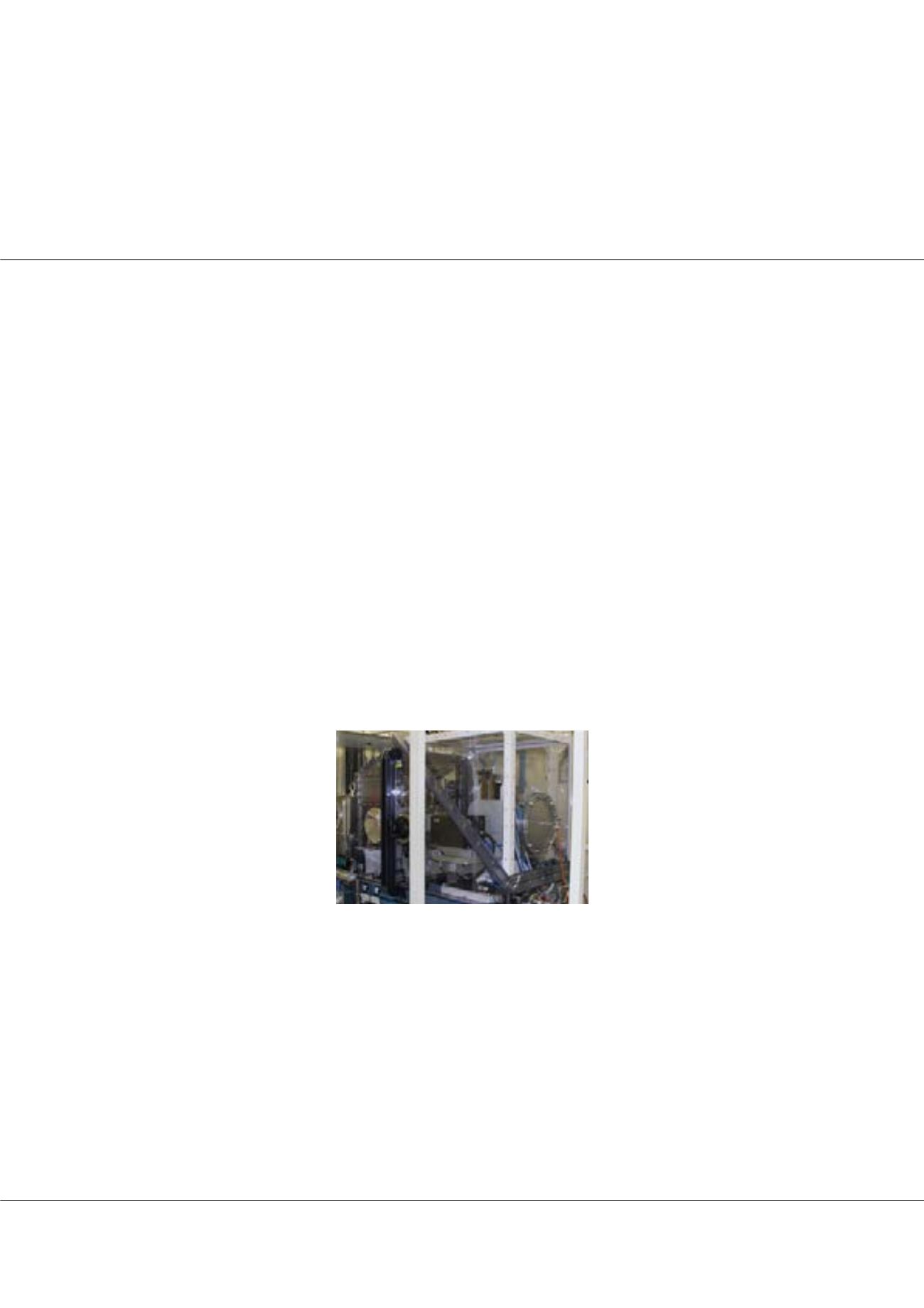
Figure1:
BL-1A of Photon Factory dedicated to the native SAD phasing