


 71 / 103

Page 109
Notes:
conferenceseries
.com
Volume 10, Issue 8 (Suppl)
J Proteomics Bioinform, an open access journal
ISSN: 0974-276X
Structural Biology 2017
September 18-20, 2017
9
th
International Conference on
Structural Biology
September 18-20, 2017 Zurich, Switzerland
Carol A Heckman, J Proteomics Bioinform 2017, 10:8(Suppl)
DOI: 10.4172/0974-276X-C1-0100
Structural aspects of cell signaling
Carol A Heckman
Bowling Green State University, USA
Statement of the Problem:
Endpoints such as adhesion and motility have been used to infer the function of a protein in
cells. These endpoints are unsatisfactory, because a protein can be recruited to different substructures and promote different
outcomes in such structures. By defining meaningful endpoints, it is possible to identify a protein’s contribution to several
different patterns of cell organization and thereby address major problems in biology.
Methodology &Theoretical Orientation:
We developed an unbiased method of classifying and quantifying features of fixed,
adherent epithelial cells. Primary data, consisting of 102 measures of contour geometry, curvature, relationship to derived
model figures, etc., were used to calculate 20 latent factors representing cell features. Factors detect structure by recognizing
the relationships between variables. Cells from experiments are classified according to each factor by summing the factor
loadings. Filopodia (factor 4) accounted for a larger proportion of cancer-related variance than any other feature. Filopodia
are the sensory appendages that are relied on when cells distinguish their more and less adhesive sides. The protrusion defined
as factor 7 represented neurites. Even when small, neurites differed from lamellipodia (factor 5). Several factors contribute to
ruffling.
Results:
Filopodia are down-regulated by three isoforms of protein kinase C (PKC). The effect of PKC ε, a known oncogene, on
filopodia is only observed after tumor promoter treatment. The effect is in part due to a PKC ε-mediated increase in ruffling.
In cells not treated with tumor promoter, filopodia are down-regulated by isoforms
α
and
ή
. PKC
α
has contrary effects in
promoter-treated cells, where it conserves filopodia by suppressing ruffling activity. Activated PKC
α
may promote filopodia.
These activities are consistent with the concept that filopodia are implicated in cell homeostasis. By regulating the prevalence
of filopodia, PKC can regulate the way cells react to their surroundings.
Biography
Carol A Heckman is an expert on Preneoplasia and image analysis and has developed optical and computational methods suitable for cell feature analysis.
Applying these methods, she showed how to deconstruct the cell phenotype and find the features related to oncogenic transformation. About half of the 20 features
are useful in distinguishing the phenotypes of normal and cancerous epithelial cells. She has published numerous papers on these topics. The cell features form
the basis of an assay to flag chemicals of interest for drug development and identify diseases that can be targeted productively by existing drugs.
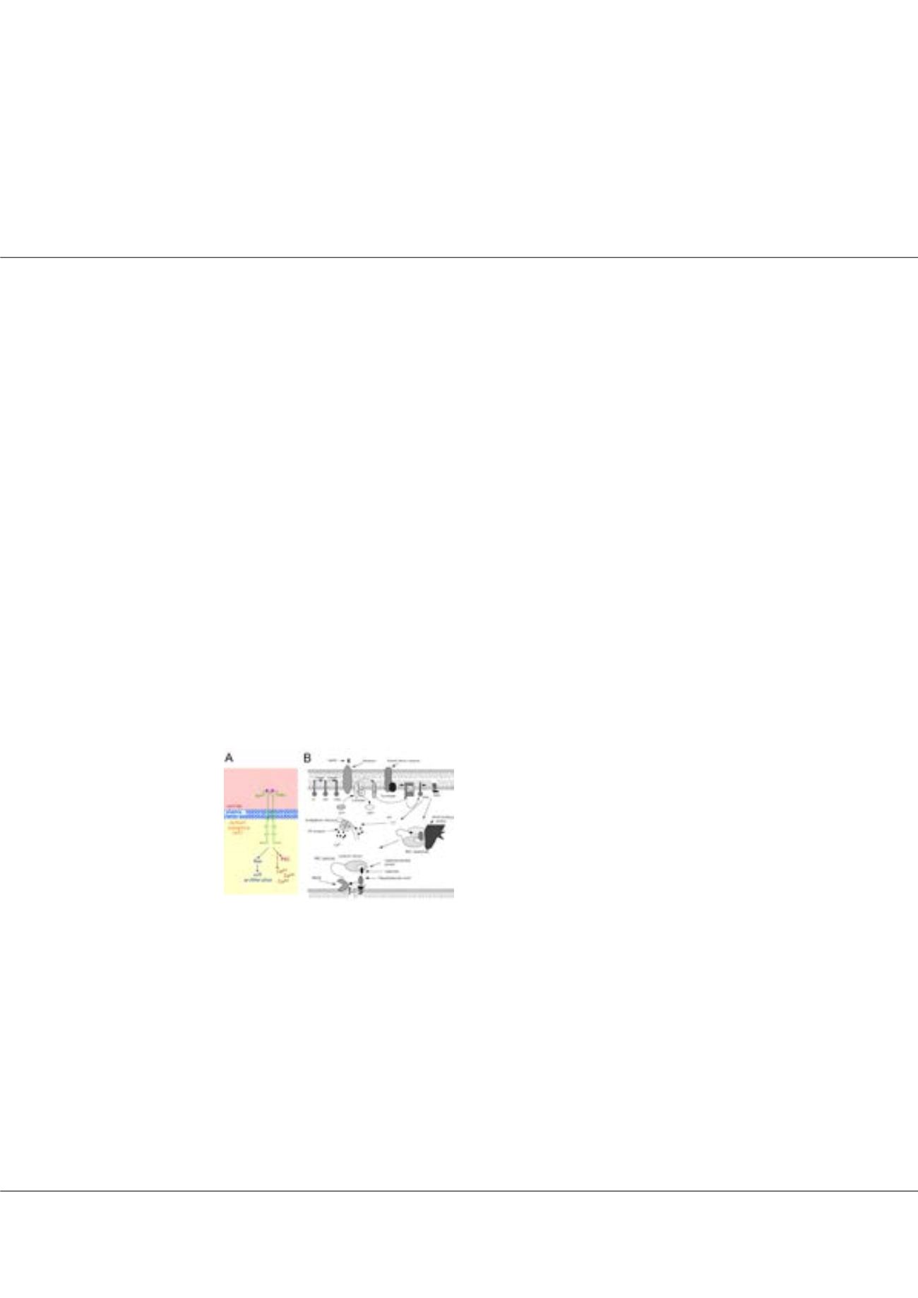
heckman@bgsu.eduFigure1:
PKC signaling. A) PKC signals
downstream of growth factor receptors. B) PKC
is also activated by G protein-coupled receptors.
When activated, it finds most of its substrates in
the vicinity of membranes.