


 51 / 103

Volume 10, Issue 8 (Suppl)
J Proteomics Bioinform, an open access journal
ISSN: 0974-276X
Structural Biology 2017
September 18- 20, 2017
Page 88
conference
series
.com
9
th
International Conference on
Structural Biology
September 18-20, 2017 Zurich, Switzerland
Carsten Mim, J Proteomics Bioinform 2017, 10:8(Suppl)
DOI: 10.4172/0974-276X-C1-0100
When structure leads to function: Protein complexes at the membrane in endocytosis
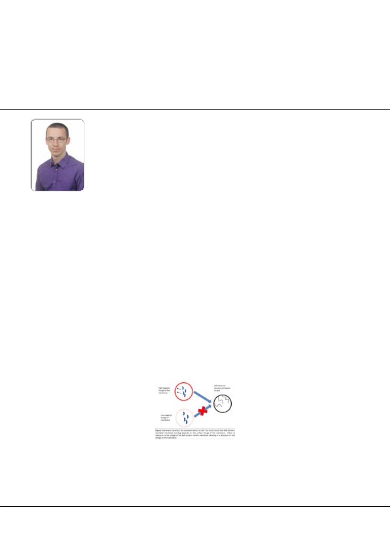
Statement of the Problem:
The cell bends membranes to generate membrane structures, like the t-tubules in muscles. Bin1/
Amphiphysin/Rvs domain proteins are part of the membrane bending machinery and are found in widespread phenomena
like endocytosis or cell motility. BAR domain proteins can assemble spontaneously
in vitro
as well as
in vivo
. Which factors
regulate the assembly, the membrane tension is a well-studied regulator. In contrast, the role of the membrane composition, as
an initiator of membrane bending, is poorly understood.
Methodology & Theoretical Orientation:
For this study, we collected electron micrographs to document the membrane
bending activity of the BAR protein Bin1. We probed the electrostatic interactions between Bin1 and the membrane by
changing the surface charge of the membrane, the ionic strength of the assay and using disease relevant mutants, where the
positive charge (K35N) and the negative charge (D151N) are eliminated. The electrostatic interactions between Bin1 and
artificial membranes were evaluated by liposome sedimentation. To test how the findings, translate into living cells, we assayed
the phenotype of membrane bending-deficient Bin1 mutants in cells that have elevated or reduced levels of negatively charged
lipids.
Findings:
Our simple, artificial system could reproduce the complex membrane topology present in muscle cells. We focused
on the two mutants. We found that in stringent conditions for membrane bending (high ionic strength, low membrane charge)
the mutants showed disproportional lower bending activity. These finding were confirmed
in vivo
. We could rescue to mutant
phenotype by increasing the membrane surface charge. Conversely, we induced a mutant phenotype in wt Bin1 by lowering
the membrane surface charge.
Conclusion & Significance:
We established the membrane charge as a novel regulator of membrane tubulation. We speculate
that rapid phosphorylation and dephosphorylation of phosphoinositols can act as a switch for induction of membrane bending.
Biography
Carsten Mim has a longstanding interest in membrane and membrane-associated proteins throughout his career. As an experienced Electrophysiologist, he characterized
the glutamate transporter EAAT3 and EAAT4. The kinetics of EAAT4 differ from other glutamate transporters, by a voltage sensitive step that slows the turnover rate at
hyperpolarized membrane potentials. Further, recorded transient and steady state currents at different temperatures showed that the binding of glutamate is enthalpy-driv-
en unlike the binding of Na+. To visualize membrane: protein complexes, he turned to electron microscopy. His work on the Bin/Amphyphysin/Rvs domain (BAR) protein
endophilin in complex with the bilayer resulted in the unexpected discovery that the stability and dynamics of endophilin scaffolds entirely depend on non-specific inter-
actions between amphipathic helices in the bilayer. His findings also provided a first structurally motivated hypothesis how BAR-scaffolds selectively recruit downstream
interaction partners through a steric selection mechanism.
carsten.mim@sth.kth.seCarsten Mim
KTH Royal Institute of Technology, Sweden