


 102 / 103

Page 141
Notes:
conferenceseries
.com
Volume 10, Issue 8 (Suppl)
J Proteomics Bioinform, an open access journal
ISSN: 0974-276X
Structural Biology 2017
September 18-20, 2017
9
th
International Conference on
Structural Biology
September 18-20, 2017 Zurich, Switzerland
Nadia L Opara et al., J Proteomics Bioinform 2017, 10:8(Suppl)
DOI: 10.4172/0974-276X-C1-0100
Delivery methods for free electron lasers: Direct protein crystallization on solid supports economizes
sample consumption in serial femtosecond crystallography
Nadia L Opara
1,2
, Isabelle Martiel
1
, Stefan A Arnold
2
, Thomas Braun
2
, Henning Stahlberg
2
and
Celestino Padeste
1,2
1
Paul Scherrer Institute, Switzerland
2
University of Basel, Switzerland
C
lassical crystallography methods based on synchrotrons usually require crystals of relatively large dimensions, i.e. above 5
micrometres. The recent availability of X-ray free electron laser sources providing femtosecond X-ray pulses of ultrahigh
brightness facilitate the investigation of nanocrystals. However, in this case data collection must be performed in the mode of
the serial crystallography in so-called diffraction-before-destruction regime because the probed area of the sample is destroyed
after the interaction with ultra-intense radiation. As thousands of crystals must be provided sequentially to the XFEL beam,
selection of an efficient sample delivery system is crucial to minimize protein consumption during data collection. Delivery
methods applied so far include steady streaming liquid jets of the crystal suspension. The application of more viscous media like
lipidic cubic phase, agarose or hyaluronic acid matrices has also been demonstrated. However, all these methods use significant
amounts of the precious protein, which cannot be recovered even if not directly probed. Recent developments of drop of
demand methods or fixed targets allow overcoming this problem. But still, handling of the fragile crystals should be gentle or
at best avoided. Microfabricated silicon chips with ultrathin Si3N4 membranes provide the possibility to regularly position
crystals on precisely defined spots by direct crystallization using classical vapor diffusion method. The sample consumption is
minimal since crystal growth takes place in nanolitre volume cavities. No additional sample transfer is needed, because X-rays
are probing the crystals at the spot where they grew on the X-ray-transparent ultrathin amorphous silicon nitride membranes.
Assembly with a second chip to form a hermetically sealed sandwich protects specimens from dehydration and facilitates
in
situ
diffraction data collection at room temperature, as demonstrated in a synchrotron experiment providing high-resolution
patterns.
Biography
Nadia L Opara joined the CINA group at the Biozentrum University of Basel and the LMN at PSI in Switzerland in 2014 in the frame of SNI PhD school program, to
work on a project aiming at improving sample preparation methods for XFEL-based protein nano-crystallography. Beforehand she completed her Bachelor studies
in chemistry and Master program in Molecular Biotechnology at the Lodz University of Technology in Poland.
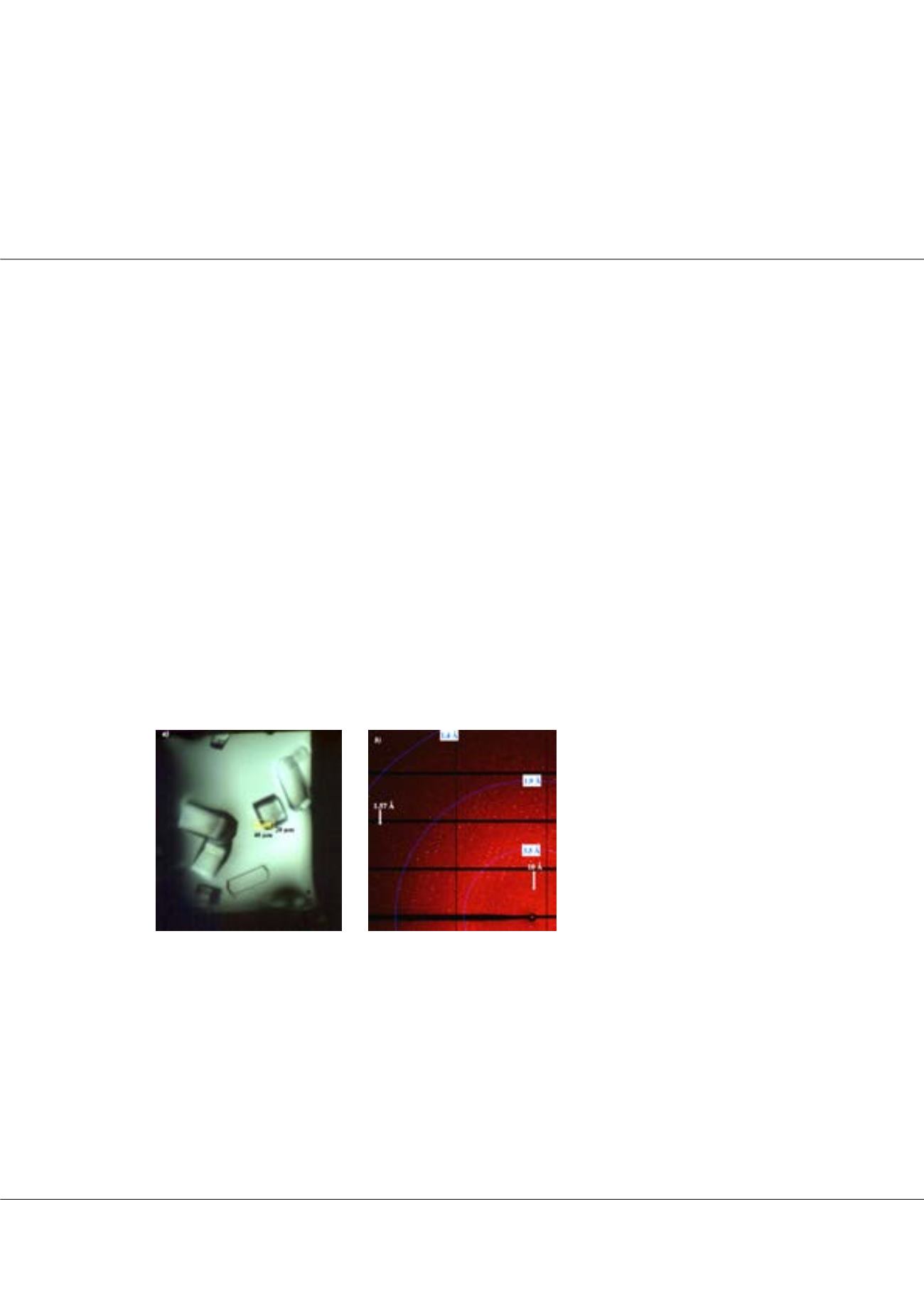
nadia.opara@gmail.comFigure1:
a) Lysozyme crystals enclosed
in-between silicon nitride membranes
and mounted on a synchrotron beamline
goniometer. b) High resolution diffraction
pattern obtained by in situ exposure with
synchrotron radiation from the crystal as
indicated in (a).