
conferenceseries
.com
Volume 6, Issue 4 (Suppl)
J Spine, an open access journal
ISSN: 2165-7939
Page 50
July 24-26, 2017 Rome, Italy
&
Spine and Spinal Disorders
2
nd
International Conference on
Neurology and Neuromuscular Diseases
6
th
International Conference on
CO-ORGANIZED EVENT
Early detection of glioma sphere xenografts models: a diffusion MR study at 14.1 T
Paola Porcari
Newcastle University, UK
Statement of the Problem:
Diffuse gliomas (WHO grade II to IV) are the most common primary brain tumours in humans, typically
associate with a severe prognosis and their diffuse infiltration into the surrounding normal brain precludes complete resection and
they all eventually recur, usually having progressed to a more aggressive tumour. The infiltrative part, which is “invisible” using
conventional T1 and T2-weighted MRI is difficult to target with treatment. We investigated whether diffusion MRI might be a useful
method to detect the microstructural changes induced in the normal brain by the slow infiltration of glioma sphere cells. Additionally,
localized proton MR spectroscopy of lesions and immunohistochemical assessment were compared with imaging results.
Methodology:
LN-2669GS and LN-2540GS (3) orthotopic glioma xenografts were induced in athymic mice. Gliomas were monitored
at 14.1T. MRI-protocol included T2-weighted turbo-spin-echo images, diffusion weighted imaging and diffusion tensor imaging, both
acquired using the pulse-gradient-stimulated-echo sequence (4) (Δ/δ=80/4ms), which allows exploring long diffusion times. Mean
values of diffusion indices were calculated in tumours and in the corresponding brain regions of controls. Proton spectra of gliomas
were acquired during the last MRI session. Finally, mice were sacrificed and sections stained underwent immunohistochemical
assessment.
Findings:
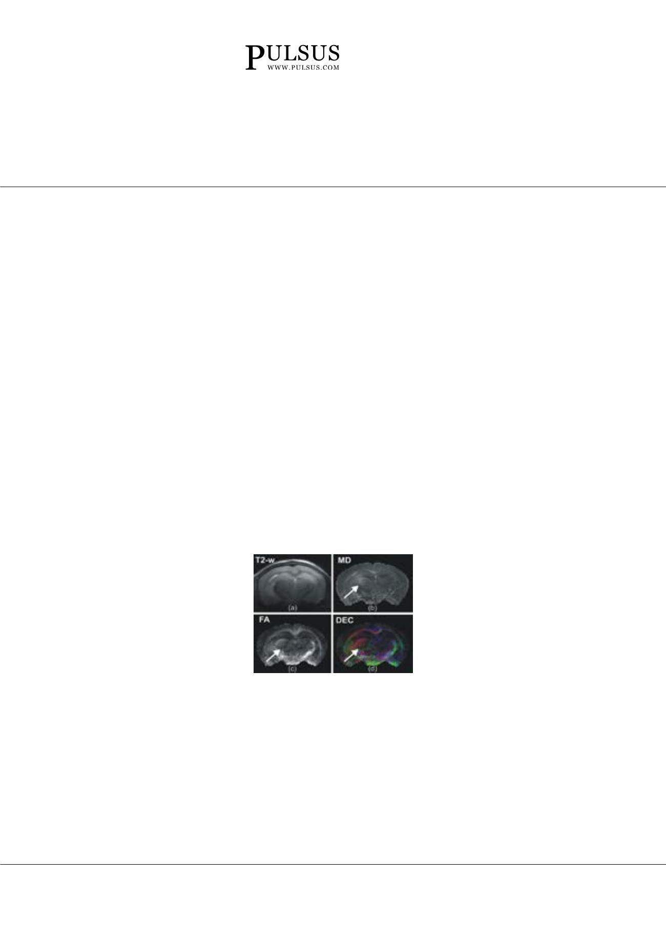
Compared with T2-weighted images, tumours were properly identified in their early stage of growth using diffusion
MRI (Figure 1) at a moderately long diffusion time. MRI results were confirmed by localized proton MR spectroscopy and
immunohistochemistry. First evidence of tumour presence was revealed for both glioma models three months after tumour
implantation, while no necrosis or haemorrhage were detected by MRI or histology.
Conclusion & Significance:
This study demonstrates that diffusion MRI is a useful method to detect and follow slowly growing,
diffuse infiltrative tumours in mouse brain, providing a potential imaging biomarker for early detection of diffuse infiltrative gliomas
in humans.
Figure: In vivo MRI of LN-2540GS glioma sphere xenograft, five months after injection of the cells. T2-weighted (a) MD (b),
FA (c) and FA-modulated DEC maps of a coronal slice from the mouse brain with the LN-2540GS xenograft NCH-1365. MD,
FA and DEC maps, computed after DTI reconstruction, show a lesion (indicated by arrow).
Biography
Paola Porcari has her expertise in high field MRI imaging of small animals. She is particularly interested in the development of new MRI methods to better
understand central nervous system diseases and neuromuscular disorders. During her current work, she developed diffusion methods based on the stimulated
echo sequence (STE-DWI) to investigate whether diffusion MRI can be a powerful technique for investigating muscle pathology. Previously she worked extensively
on glioma model applying different MRI techniques, such as, diffusion MRI and multi-nuclear NMR, especially 19F MRI/MRS to evaluate different aspect of the
disease.
paola.porcari@ncl.ac.ukPaola Porcari, J Spine 2017, 6:4(Suppl)
DOI: 10.4172/2165-7939-C1-005


