


 9 / 21

Page 31
Notes:
conferenceseries
.com
Volume 6, Issue 4(Suppl)
OMICS J Radiol, an open access journal
ISSN: 2167-7964
Medical Imaging and Clinical Research 2017
September 11-12, 2017
September 11-12, 2017 | Paris, France
2
nd
World Congress on
Medical Imaging and Clinical Research
Dynamics of folliculogenesis–sonographic assessment and applications in infertility management
Monica Kansal
Jaypee hospital, India
T
rans-vaginal sonography along with colour Doppler is the gold standard investigation in assessment of gynaecological and
reproductive disorders in females. Besides exclusion of uterine, endometrial and tubal causes, sonography provides non-
invasive tool for monitoring individual follicles during menstrual cycle and response to ovarian stimulation. This paper describes
various uses of ultrasound in assisted reproductive techniques as the principal non-invasive modality for evaluation of key process
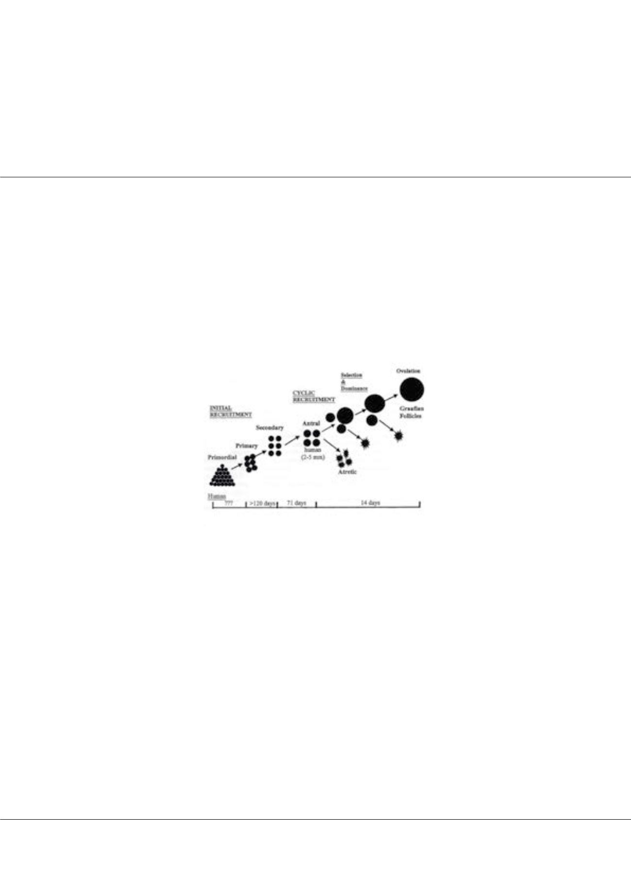
of ovarian function – the process of folliculogenesis. Folliculogenesis refers to all phases that a primordial germ cell should pass to
become mature healthy oocyte that is subsequently fertilized. It is a constant process that starts in embryogenic period and ends with
the disappearance of last functional follicle in the period of menopause. Recognising the quality of follicle, its growth pattern and
vascularity has a prognostic value for outcome of assisted reproduction techniques.
Figure 1:
Folliculogenesis - Process of follicle recruitment and development.
Biography
Monica Kansal has been practising Radiology at eminent hospital and educational institutions from last nine years with special interest in Women’s imaging.
mk_mamc@yahoo.co.inMonica Kansal, OMICS J Radiol 2017, 6:4(Suppl)
DOI: 10.4172/2167-7964-C1-012