


 20 / 21

Page 44
Notes:
conferenceseries
.com
Volume 6, Issue 4(Suppl)
OMICS J Radiol, an open access journal
ISSN: 2167-7964
Medical Imaging and Clinical Research 2017
September 11-12, 2017
September 11-12, 2017 | Paris, France
2
nd
World Congress on
Medical Imaging and Clinical Research
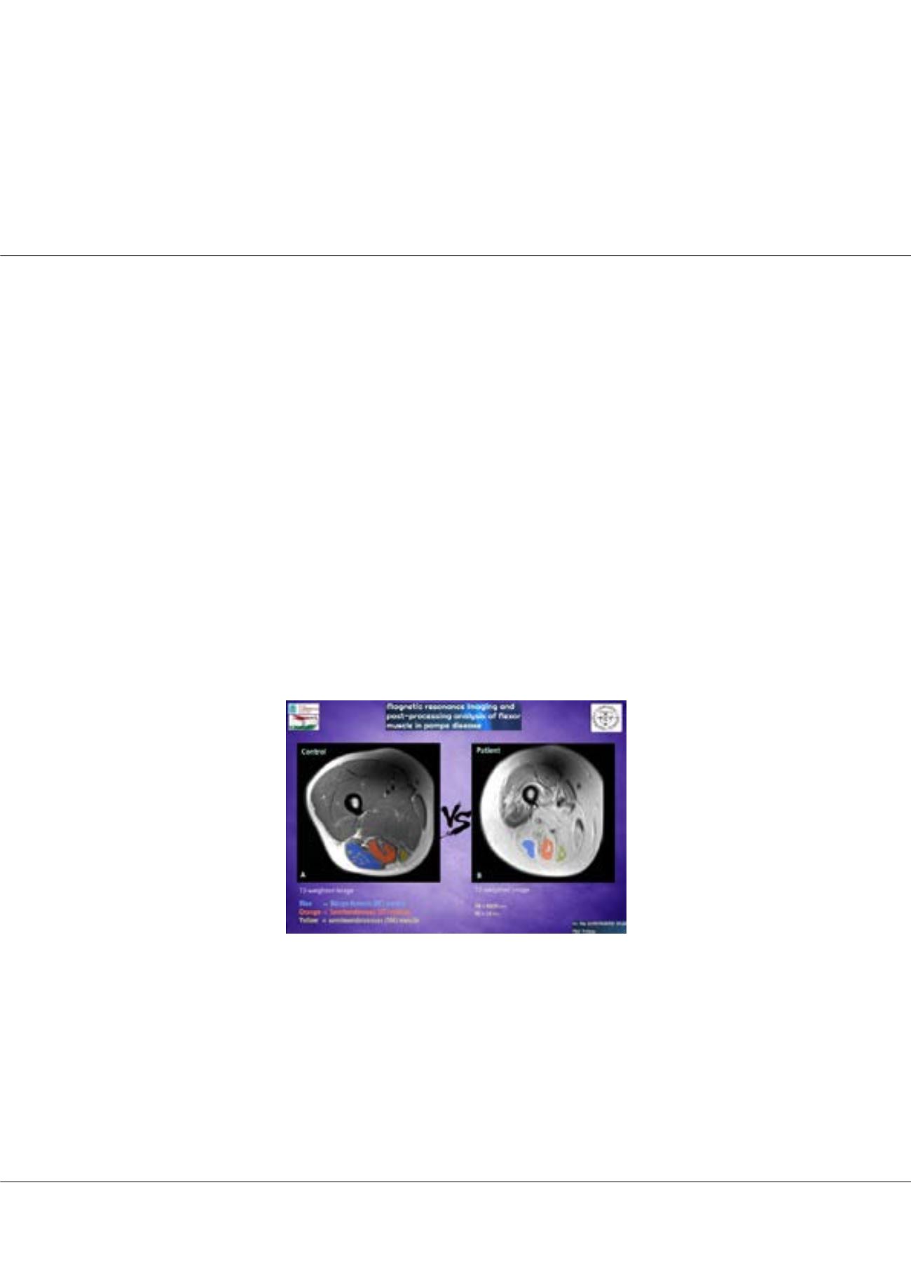
Magnetic resonance imaging and post-processing analysis of flexor muscle in Pompe disease
Ala Khasawneh
University of Pécs, Hungary
P
ompe disease is a rare multisystem genetic disorder that characterized by a deficiency of the lysosomal enzyme acid alpha-
glucosidase and cytoplasmic glycogen accumulation causing damage that leads to muscle weakness. This study aim is to evaluate
the muscle MRI pattern of twelve adults with late onset Pompe disease and twelve sex- and age-matched healthy controls (Age
range 19-59) for feature extraction which will be used to identify and classify functioning and non-functioning muscles. A training
procedure was implemented using an Exercise Dynamometer device to stimulate the muscle for maximal contraction, MRI images
data was used to compare the three flexor muscles in the lower limb function. MRI images data of biceps femoris (BF) muscle,
Semitendinosus (ST) muscle, semimembranosus (SM) muscle before and after exercise (base, 30 min, 24 hours) was measured. We
performed and quantified T2 relaxation data of flexor muscles, and all data analyzed using repeated measure ANOVA to compare
within related groups of the independent variable time (Base, 30M, 24H). According to our results, the significantly lower T2 value
in the ST muscle of controls was observed (base=43ms, 30min=48ms, 24h=43ms; P < 0.05), but the change in SM muscle and BF
muscle were not significant. In patients, we detected significantly higher T2 value in SM muscle evolve over time (base=129ms,
30min=132ms, 24h=128ms; P < 0.05) compared to the controls, but ST muscle neither BF muscle doesn't show significant change. As
a conclusion, we can say that in Pompe patients the SM muscle can only react to the exercise apparently and shows us an activity in
affected muscle cells, compared to the BF and ST muscle not shown any activity, that’s mean perhaps the Pompe disease change the
muscle cells structure to interact to the exercise.
Biography
Ala Khasawneh is a Jordanian Doctor. He completed his Diploma in General Medicine (MD) and has been awarded the qualification of a Physician and title of
Doctor of Medicine from National Pirogov Memorial Medical University. He worked in Basma Hospital, Jordan. Currently, he is a PhD fellow in Diagnostic Medical
Imaging in Hungary. As a Doctor, his main interest is to create new pathways for improving health care.
alaakhalel@aol.comAla Khasawneh, OMICS J Radiol 2017, 6:4(Suppl)
DOI: 10.4172/2167-7964-C1-012