
Page 63
Notes:
conferenceseries
.com
Volume 3
Diagnostic Pathology: Open Access
ISSN: 2476-2024
Laboratory Medicine 2018
June 25-26, 2018
June 25-26, 2018 | Berlin, Germany
13
th
International Conference on
Laboratory Medicine & Pathology
Glioma Tumor Segmentation Using Deep Cascaded – 3D Convolutional Neural Networks
Wiem Takrouni
and
Ali Douik
University of Sousse, Tunisia
G
lioma is a brain tumor, mainly dangerous form of cancer which starts in the gluey supportive cells (glial cells) that encircle
nerve cells and help them function. Glioma tumor brain structure segmentation in non-invasive magnetic resonance
images (MRI) has enticed the interest of the research community for a long time because of morphological changes in these
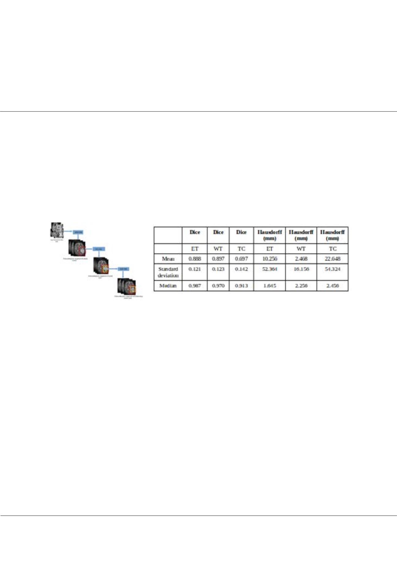
structures. In this poster, we propose a deep cascaded 3Dconvolutional neural network (DC-3DCNN) basedmethod to segment
glioma lesions from multi-contrast MR images. DC-3DCNN was evaluated on a challenge dataset, where the performance of
our method is also compared with different current publicly available state-of-the-art lesion segmentation methods.
Biography
I’m Wiem Takrouni from Tunisia, i’m a Phd Student in computer vision and Medical imaging in the (NOCCS Laboratory, National School of Engineering of Sousse,
University of Sousse, Tunisia). I have three publications paper conferences: two publications in smart vehicule with deep learning and a publication in tumor
reconstruction in medical imaging.
Suhi14@yahoo.com takrouni.wiem@gmail.comWiem Takrouni et al., Diagn Pathol Open 2018, Volume 3
DOI: 10.4172/2476-2024-C1-003
Table1:Dice and Hausdorff measurements of the DC-
3DCNN method on BraTS 2017 denote enhancing tumor
core, whole tumor and tumor core, respectiv
Figure1: Proposed Method


