


 13 / 37

Page 39
Volume 09
Otolaryngology: Open Access
ISSN: 2161-119X
ENT 2019
Craniofacial Surgery 2019
August 15-17, 2019
JOINT EVENT
conferenceseries
.com
August 15-17, 2019 Rome, Italy
&
3
rd
International Conference on
Craniofacial Surgery
4
th
European Otolaryngology-ENT Surgery Conference
Auricular schwannoma: A case report
Raid M. AL-Ani
and
Haidar Khudair Abd
University of Anbar, College of Medicine, Iraq
S
chwannoma is a benign tumour of schwann cells and is seldom to be seen in the auricle. In the literature, very few
cases of schwannomas originating in the pinna were reported. In this article, we described a 35-year-old female
patient who presented with right painless auricular mass which was treated by excision under general anesthesia.
The clinical and histopathologic features, the differential diagnosis, and the treatment of auricular schwannoma are
discussed.
Key words
: auricle, schwannoma, Iraq.
Introduction
: Schwannomas are slowly growing benign tumour of neuro-ectodermal origin. Schwannomas are well
known to arise from schwann cell of the branches of peripheral, cranial or autonomic nerves. They are usually
presented as a painless solitary swelling. They are affecting the head and neck in a 25-45%, where the vestibular
schwannoma is the commonest. The presentation of head and neck schwannomas depends on their location. Auricle
is a rare site of affection by schwannoma (1). The first case of external ear schwannoma was reported in 1977 (2).
When we were reviewing the literatures, only five cases of auricular schwannomas were reported in the world (1-5).
In the present article we describe a further case of auricular schwannoma.
Verocay in 1908 was the first who describe the solitary schwannoma and gave it the name of neurinoma; the name
schwannoma was assigned by Batsakis in 1974. Schwannoma is also known by other terms, such as neurinoma,
neurilemmoma, mioschwannoma, schwannoglioma, etc (6). The first case of auricular schwannoma was reported by
Fodor et al in 1977. Following this case only 4 cases were reported in the world (1-5).
Raid M. AL-Ani et al., Otolaryngol (Sunnyvale) 2019, Volume 09
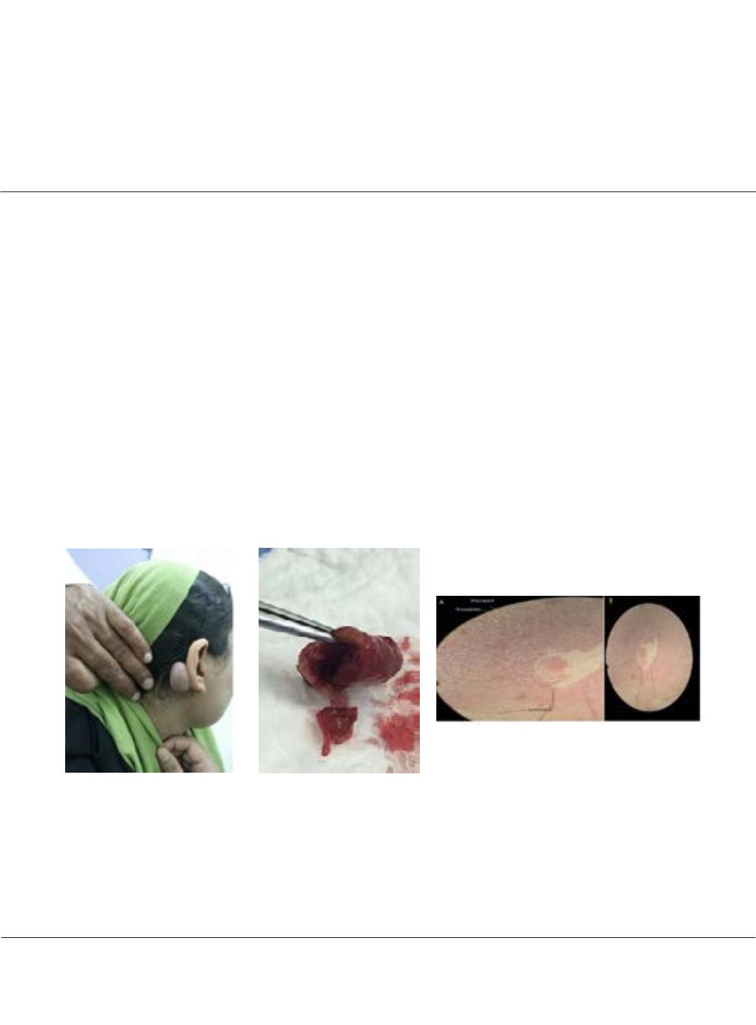
Figure 1: The patient with right
auricular mass.
Figure 2: The excised mass
contains a blood with in its cavity
and has a thick wall.
Figure 3: Microscopic section of the tumor showing areas of
compact spindle cells arrayed in a palisade pattern known as
Antoni type A, Antoni type B, Verocay bodies and an area of
hemorrhage within a cavity. (H&E staining, A: 40× and B: 4×
magnification power)