 Spanish
  Chinese
  Russian
  German
  French
  Japanese
  Portuguese
  Hindi
Hindi Our Group organises 3000+ Global Conferenceseries Events every year across USA, Europe & Asia with support from 1000 more scientific Societies and Publishes 700+ Open Access Journals which contains over 50000 eminent personalities, reputed scientists as editorial board members.
Open Access Journals gaining more Readers and Citations
700 Journals and 15,000,000 Readers Each Journal is getting 25,000+ Readers
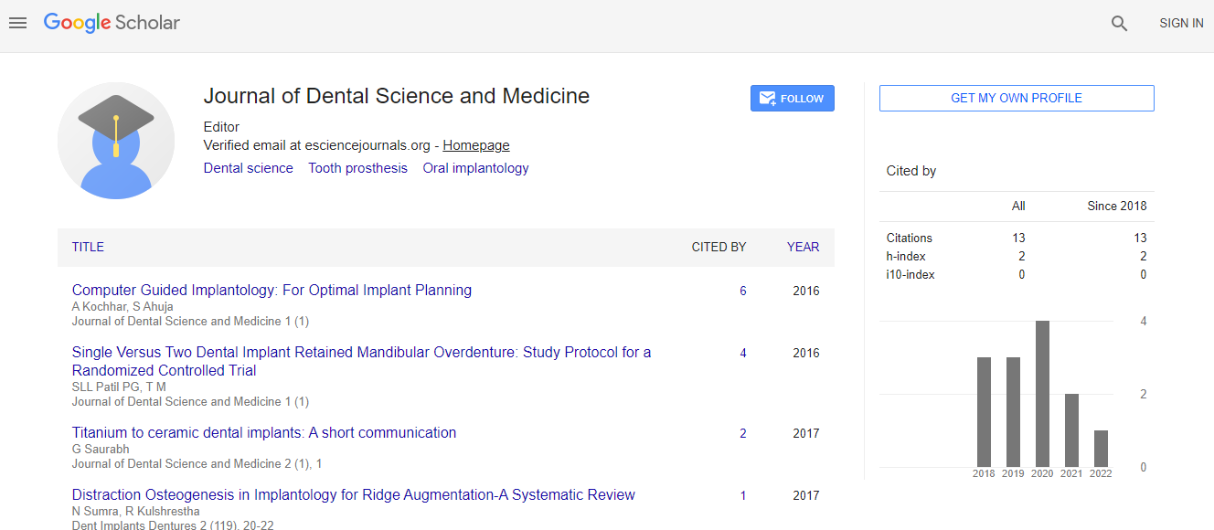
Google Scholar citation report
Citations : 13
Journal of Dental Science and Medicine received 13 citations as per Google Scholar report
Indexed In
- RefSeek
- Hamdard University
- EBSCO A-Z
- ICMJE
Useful Links
Recommended Journals
Related Subjects
Share This Page
Visibility of anatomical landmarks in the region of the mandibular third molar, a comparison between a lowdose and default protocol of CBCT
28th American World Dentistry Congress & 35th Annual World Dentistry Summit & 8th International Conference on Prosthodontics & Orthodontics
Josefine Cederhag
Malm�?¶ University, Malm�?¶, Sweden
ScientificTracks Abstracts: Dent Implants Dentures
Abstract
European commission of radiation proection (Euratom) has elaborated guidelines, SedentexCT, with the objective to promote scientific based use of dental CBCT for varios diagnostic indications e.g., mandibular third molars. Optimization of radiographic examinations is necessary for radiation protection and many countries regulate radiation delivery to patients by law. Therefore, the aim of the study was to investigate the clinical applicability of a low-dose CBCT protocol as compared to the default for pre-surgical evaluation of mandibular third molars. Forty-eight patients (62 teeth) referred for pre-surgical mandibular third molar examination was enrolled after justification for CBCT. Two CBCT scans of each tooth were made using a default protocol and a low-dose protocol (Veraviewepocs 3D F40, J Morita Corp, Kyoto, Japan). The protocols had the same tube potential (90kV) and exposure time (9.4s), but in the low-dose protocol the tube current was reduced to 2 mA instead of 5mA. Four observers evaluated the visibility of five relevant anatomical structures and relations. The subjective image quality was ranked on a 3-point scale as diagnostically acceptable, doubtful, or unacceptable. The Wilcoxon signed-rank test compared differences between the two protocols and significance was set at P â�?¤ .05. No significant differences were found between the two protocols for any observer regarding the visibility of the relationship and proximity between the roots and the mandibular canal; root morphology; and possible root resorption of the second molar. The periodontal ligament differed significantly in visibility between the two protocols (P â�?¤ .05). The four observers ranked (mean percentage) 96% of the default images and 84% of the lowdose images as diagnostically acceptable. Two observers scored the subjective image quality for the low-dose protocol images more frequently as diagnostically doubtful compared to the default protocol images. In conclusion this study indicates that a lowdose CBCT protocol with 60% reduced tube current provides, in most cases, acceptable image quality for pre-surgical assessment of mandibular third molars. Optimization of CBCT protocols should be a primary issue according to recommended guidelines.Biography
Josefine Cederhag, DDS, PhD candidate in Oral and Maxillofacial Radiology, Malmö University, Sweden. In 2007, Josefine Cederhag graduated from the Dentist School at Umeå University, Sweden. For ten years Josefine worked as a general dentist before moving on to hold a position as a clinical teacher and clinician at the Dentist School, Malmö University, Sweden.