 Spanish
  Chinese
  Russian
  German
  French
  Japanese
  Portuguese
  Hindi
Hindi Our Group organises 3000+ Global Conferenceseries Events every year across USA, Europe & Asia with support from 1000 more scientific Societies and Publishes 700+ Open Access Journals which contains over 50000 eminent personalities, reputed scientists as editorial board members.
Open Access Journals gaining more Readers and Citations
700 Journals and 15,000,000 Readers Each Journal is getting 25,000+ Readers
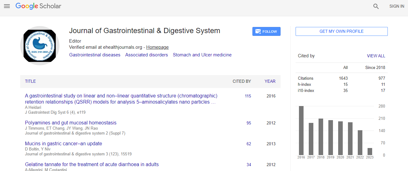
Google Scholar citation report
Citations : 1636
Journal of Gastrointestinal & Digestive System received 1636 citations as per Google Scholar report
Journal of Gastrointestinal & Digestive System peer review process verified at publons

Indexed In
- Index Copernicus
- Google Scholar
- Sherpa Romeo
- Open J Gate
- Genamics JournalSeek
- China National Knowledge Infrastructure (CNKI)
- Electronic Journals Library
- RefSeek
- Hamdard University
- EBSCO A-Z
- OCLC- WorldCat
- SWB online catalog
- Virtual Library of Biology (vifabio)
- Publons
- Geneva Foundation for Medical Education and Research
- Euro Pub
- ICMJE
Useful Links
Recommended Journals
Related Subjects
Share This Page
The mucosal loss is the critical mechanism of esophageal stricture after mucosal resection: A pilot experiment in a Porcine Model
14th International Conference on Clinical Gastroenterology and Hepatology
Bingrong Liu and Saif Ullah
The First Affiliated Hospital of Zhengzhou University, China
Posters & Accepted Abstracts: J Gastrointest Dig Syst
Abstract
Background and Aim: Esophageal stricture is a major complication of large area endoscopic mucosal resection (EMR) or endoscopic submucosal dissection (ESD). To date, the critical mechanism of esophageal stricture has not been fully elucidated. Here, we designed this experiment to explore the role of mucosal loss in esophageal stricture after mucosal resection in a porcine model. Material and Methods: Twelve swine were used for this study and randomly divided into two groups. Firstly, in all the swine, two submucosal tunnels were made of 5 cm in length and 1/3rd in width on the anterior and posterior wall of the esophageal circumference. After that, the covered mucosa was resected along the lateral edges of the tunnel in the group 1. The meanwhile covered mucosa was incised on the midline of the tunnels in the group 2. The process of stricture formation was evaluated by endoscopy after one, two and four weeks respectively. Anatomical and histological examinations were performed after euthanasia. Result: Ulcer formation was observed on endoscopy after one week. Group 1(mucosa resected) developed mild to severe esophageal stricture with dysphagia and weight loss, whereas no esophageal stricture was evident in the ones of group 2 (mucosa incised) after two and four weeks respectively. Macroscopic appearance showed severe esophageal stricture and shortening of the esophagus in the group 1 while no evident esophageal stricture and shortened esophagus was found in the group 2. Inflammations and fibrous hyperplasia of the submucosal layer were observed in both groups, on histological examination. Conclusion: The loss of esophageal mucosa might be the crucial factor for esophageal stricture after mucosal resection. Fibrosis followed by inflammation may slightly attribute toward esophageal stricture formation but is not the main mechanism of the postresection stricture. These results have significance for developing a suitable treatment for esophageal stricture.Biography
E-mail: karypton@hotmail.com