Dersleri yüzünden oldukça stresli bir ruh haline sikiş hikayeleri bürünüp özel matematik dersinden önce rahatlayabilmek için amatör pornolar kendisini yatak odasına kapatan genç adam telefonundan porno resimleri açtığı porno filmini keyifle seyir ederek yatağını mobil porno okşar ruh dinlendirici olduğunu iddia ettikleri özel sex resim bir masaj salonunda çalışan genç masör hem sağlık hem de huzur sikiş için gelip masaj yaptıracak olan kadını gördüğünde porn nutku tutulur tüm gün boyu seksi lezbiyenleri sikiş dikizleyerek onları en savunmasız anlarında fotoğraflayan azılı erkek lavaboya geçerek fotoğraflara bakıp koca yarağını keyifle okşamaya başlar

GET THE APP

The Endoscopic Diagnosis Of Early Gastric Cancer | 67015
ISSN: 2161-069X

Journal of Gastrointestinal & Digestive System
Open Access

Our Group organises 3000+ Global Conferenceseries Events every year across USA, Europe & Asia with support from 1000 more scientific Societies and Publishes 700+ Open Access Journals which contains over 50000 eminent personalities, reputed scientists as editorial board members.

Open Access Journals gaining more Readers and Citations
700 Journals and 15,000,000 Readers Each Journal is getting 25,000+ Readers
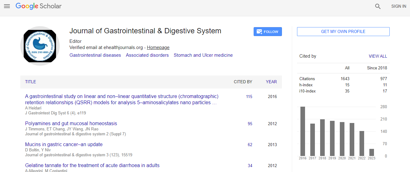
This Readership is 10 times more when compared to other Subscription Journals (Source: Google Analytics)

The endoscopic diagnosis of early gastric cancer

11th Global Gastroenterologists Meeting

Shinichi Katsuki

Otaru Ekisaikai Hospital, Japan

Posters & Accepted Abstracts: J Gastrointest Dig Syst

DOI: 10.4172/2161-069X-C1-050

Abstract
The incidence of gastric cancer in Japan is very high. Therefore, we always focus on endoscopic diagnosis of early gastric cancer. Early gastric cancer occurs in the mucous membrane and invades to the submucosal layer. The prognosis of gastric cancer depends on its stage. We detect an initial lesion of gastric cancer by observing the mucous membrane closely using an endoscope�?It leads to a higher survival rate of gastric cancer. The first step of diagnosing early gastric cancer is to find suspicious lesions by white light endoscopy. Close attention is paid to color changes of the lesions such as reddish or pale and to surface morphology changes such as elevation or depression. The second step is image enhanced endoscopy (IEE). Basically, there are two IEE methods; dyebased IEE (chromo-endoscopy) and equipment-based IEE (optical digital endoscopy). These methods enable us to recognize the demarcation line (DL) of the background mucous membrane and the cancerous lesion. The representative optical digital endoscopy is narrow band imaging system (NBI). NBI light is absorbed by hemoglobin contained in the blood vessels. With this light, we can observe blood vessels in the mucous membrane and the submucosal layer and recognize the mucous membrane microstructure. Consequently, we can diagnose early gastric cancer and identify tumor margins. The third step is magnifying observation of lesions. We can determine whether there are any irregularities of micro-vascular architecture and superficial surface structure. When we find either irregularity, the lesion is diagnosed as cancer.
Biography

Email: shinichi katsukiekisaikai@yahoo.co.jp

Top