Our Group organises 3000+ Global Conferenceseries Events every year across USA, Europe & Asia with support from 1000 more scientific Societies and Publishes 700+ Open Access Journals which contains over 50000 eminent personalities, reputed scientists as editorial board members.
Open Access Journals gaining more Readers and Citations
700 Journals and 15,000,000 Readers Each Journal is getting 25,000+ Readers
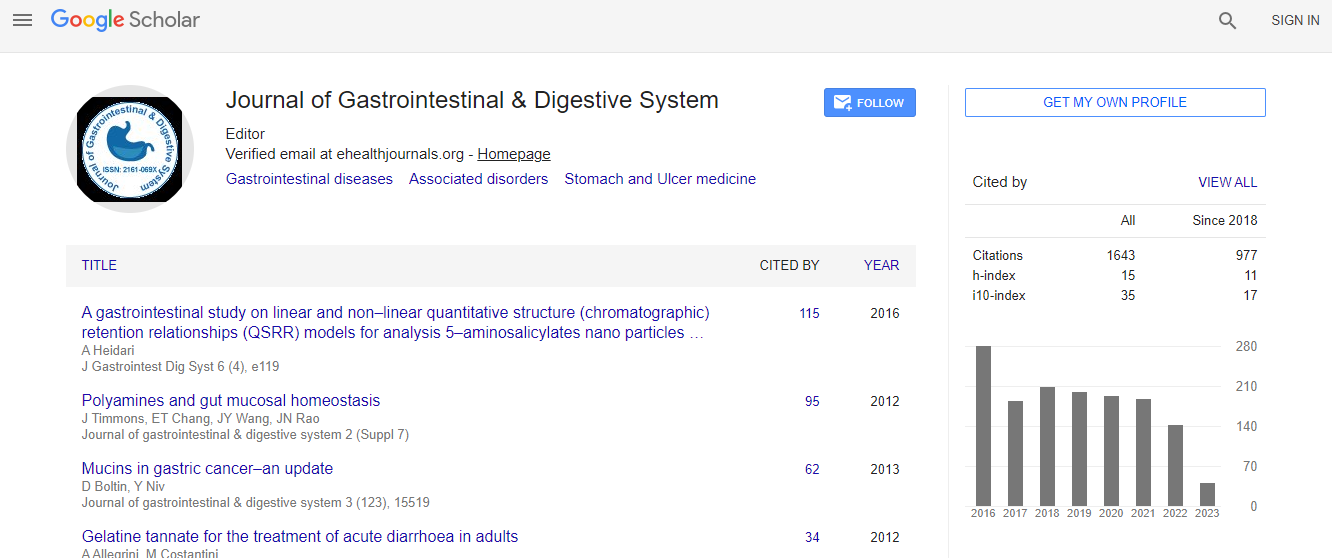
Google Scholar citation report
Citations : 2091
Journal of Gastrointestinal & Digestive System received 2091 citations as per Google Scholar report
Journal of Gastrointestinal & Digestive System peer review process verified at publons
Indexed In
- Index Copernicus
- Google Scholar
- Sherpa Romeo
- Open J Gate
- Genamics JournalSeek
- China National Knowledge Infrastructure (CNKI)
- Electronic Journals Library
- RefSeek
- Hamdard University
- EBSCO A-Z
- OCLC- WorldCat
- SWB online catalog
- Virtual Library of Biology (vifabio)
- Publons
- Geneva Foundation for Medical Education and Research
- Euro Pub
- ICMJE
Useful Links
Recommended Journals
Related Subjects
Share This Page
Technological developments in the quantitative visualization of pelvic floor function using imaging and biomechanical devices
World Congress on Gastroenterology & Urology
Christos E Constantinou
Key Note Forum: J Gastrointest Digest Sys
Abstract
The muscles associated with the pelvic floor function contribute to multiple function during a lifetime. These vary from conception to birth/delivery to urinary and bowel continence and respond to voluntary and reflex stimuli. Ultrasound imaging and image analysis enables the visualization of movement of important anatomical structures such as the bladder, vagina, and urethra. The relative displacement of such organs to incontinence producing events provides an indication of their attachment within the abdomen. Such displacement can be used to analyze the superficial pelvic floor function because it is affected by the ischiocavernous muscle, the bulbocavernosus muscle, and the perineal membrane containing muscles surrounding the vagina and the urethra. Analysis of sequences of ultrasound images generates biomechanical parameters such as relative displacement, velocity and acceleration of movement of relevant organs. However the force generated by the various pelvic floor muscles cannot be derived directly from imaging. To enable the biomechanical evaluation of muscle strength a novel vaginal probe was constructed and used to measure the force of voluntary passive, and active displacement as well reflex response at various positions within the vagina. Data were obtained from healthy volunteers as well as incontinent subjects and those with pain. Presentation illustrates the characteristic response of these subjects and integrates the data generated using the two different modalities. The visualization approach enables the direct evaluation of the differences not only between healthy and incontinent function but also aids in the evaluation of the coordinated voluntarily and involuntarily reflex contractions that responds timely to changes in intraabdominal activity. Consequent dysfunction that may produce disorders, which such as incontinence, lower urinary tract symptoms and pain can be quantified. Furthermore, image analysis of ultrasound visualizations is advantageous in exploring relative dynamic anatomy by providing simultaneously considerable amount of dynamic data of different pelvic structures that cannot be visually assimilated in real time by the observer, particularly during fast events such as coughs.Biography
Chris Constantinou has completed his Ph.D at Stanford University and continued as faculty in the department of Urology. He is now the principal investigator of an NIH project involving the evaluation of pelvic floor function at the VA Medical Center in Palo Alto at the Spinal cord injury center. He has published more than 136 papers and chapters in peer reviewed journals, is Editor in Chief of the Open Journal of Obstetrics and Gynecology and is serving on the editorial board member of many other journals.

 Spanish
  Chinese
  Russian
  German
  French
  Japanese
  Portuguese
  Hindi
Hindi 
