Our Group organises 3000+ Global Conferenceseries Events every year across USA, Europe & Asia with support from 1000 more scientific Societies and Publishes 700+ Open Access Journals which contains over 50000 eminent personalities, reputed scientists as editorial board members.
Open Access Journals gaining more Readers and Citations
700 Journals and 15,000,000 Readers Each Journal is getting 25,000+ Readers
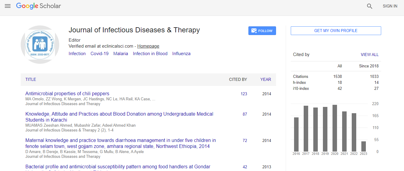
Google Scholar citation report
Citations : 1529
Journal of Infectious Diseases & Therapy received 1529 citations as per Google Scholar report
Indexed In
- Index Copernicus
- Google Scholar
- Open J Gate
- RefSeek
- Hamdard University
- EBSCO A-Z
- OCLC- WorldCat
- Publons
- Euro Pub
- ICMJE
Useful Links
Recommended Journals
Related Subjects
Share This Page
Skin alterations: Prolonged use of steroids by dermoscopy
International Conference on Infectious Diseases, Diagnostic Microbiology & Dermatologists Summit on Skin Infections
Alin Laurentiu Tatu
Dunarea de Jos University, Romania
Posters & Accepted Abstracts: J Infect Dis Ther
Abstract
Objectives: The aims of this study were to investigate if the skin alterations after prolonged use of steroids are highlighted by dermoscopy. Methods: Patients with variable facial lesions included as (SIFD) after prolonged use of topical steroids more than nine months minimum twice weekly were examined clinically and by Dermoscopy. Results: All patients showed telangiectasias (100%) and dermoscopy revealed linear, tortuous and polygonal vessels.72% of the patients had dermoscopic features for Demodex folliculorum-follicular plugs and Demodex tails. All the 29% patients with clinical spinulosus had Demodex dermoscopic features.76% of the patients had clinically visible pustules but by dermoscopy the tiny infraclinical pustules could be seen better and earlier. 77% of the patients had visible erythema on the face and by dermoscopy all they had red diffuse areas. The white hairs derived from hypertrichosis were observed at 13% with the naked eye and at 43% by dermoscopy. The atrophy was clinically visible at 12% patients as a severe skin thinning but dermoscopy revealed also atrophic areas at another 2 patients as white structure less areas or patches between vessels. The patients with dermoscopic atrophy were using mometasonefuroat and clobetasol propionate. Conclusions: The dermoscopic particularity of steroid induced rosacea is the association of white intervascular structure less patches or areas as a sign of the atrophy and also the early detection of hypertrichosis. Limitations: The small number of the patients may not accurately reflect the percent of dermoscopic findings.Biography
Email: dralin_tatu@yahoo.com

 Spanish
  Chinese
  Russian
  German
  French
  Japanese
  Portuguese
  Hindi
Hindi 