 Spanish
  Chinese
  Russian
  German
  French
  Japanese
  Portuguese
  Hindi
Hindi Our Group organises 3000+ Global Conferenceseries Events every year across USA, Europe & Asia with support from 1000 more scientific Societies and Publishes 700+ Open Access Journals which contains over 50000 eminent personalities, reputed scientists as editorial board members.
Open Access Journals gaining more Readers and Citations
700 Journals and 15,000,000 Readers Each Journal is getting 25,000+ Readers
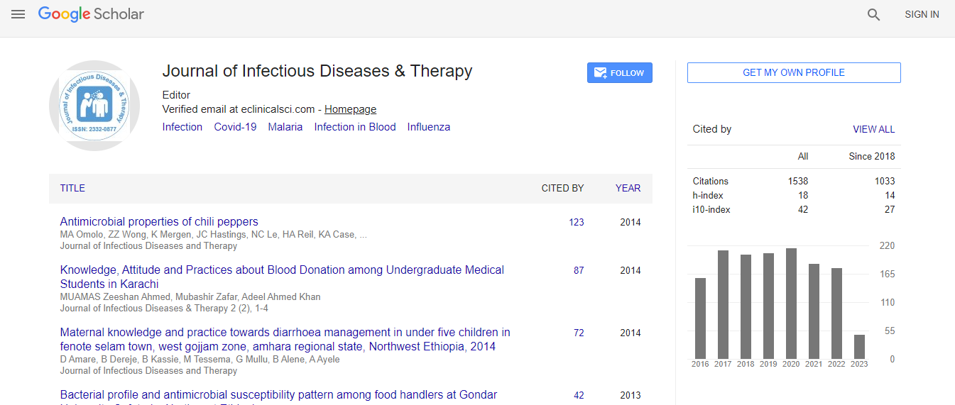
Google Scholar citation report
Citations : 1529
Journal of Infectious Diseases & Therapy received 1529 citations as per Google Scholar report
Indexed In
- Index Copernicus
- Google Scholar
- Open J Gate
- RefSeek
- Hamdard University
- EBSCO A-Z
- OCLC- WorldCat
- Publons
- Euro Pub
- ICMJE
Useful Links
Recommended Journals
Related Subjects
Share This Page
Recurrent cholangitis associated with biliary sludge and Phrygian cap anomaly diagnosed by magnetic resonance imaging and magnetic resonance cholangiopancreatography despite normal ultrasound and computed tomography
6th Euro-Global Conference on Infectious Diseases
Basaranoglu Metin
Bezmialem Vakif University Faculty Hospital, Istanbul, Turkey
Posters & Accepted Abstracts: J Infect Dis Ther