 Spanish
  Chinese
  Russian
  German
  French
  Japanese
  Portuguese
  Hindi
Hindi Our Group organises 3000+ Global Conferenceseries Events every year across USA, Europe & Asia with support from 1000 more scientific Societies and Publishes 700+ Open Access Journals which contains over 50000 eminent personalities, reputed scientists as editorial board members.
Open Access Journals gaining more Readers and Citations
700 Journals and 15,000,000 Readers Each Journal is getting 25,000+ Readers
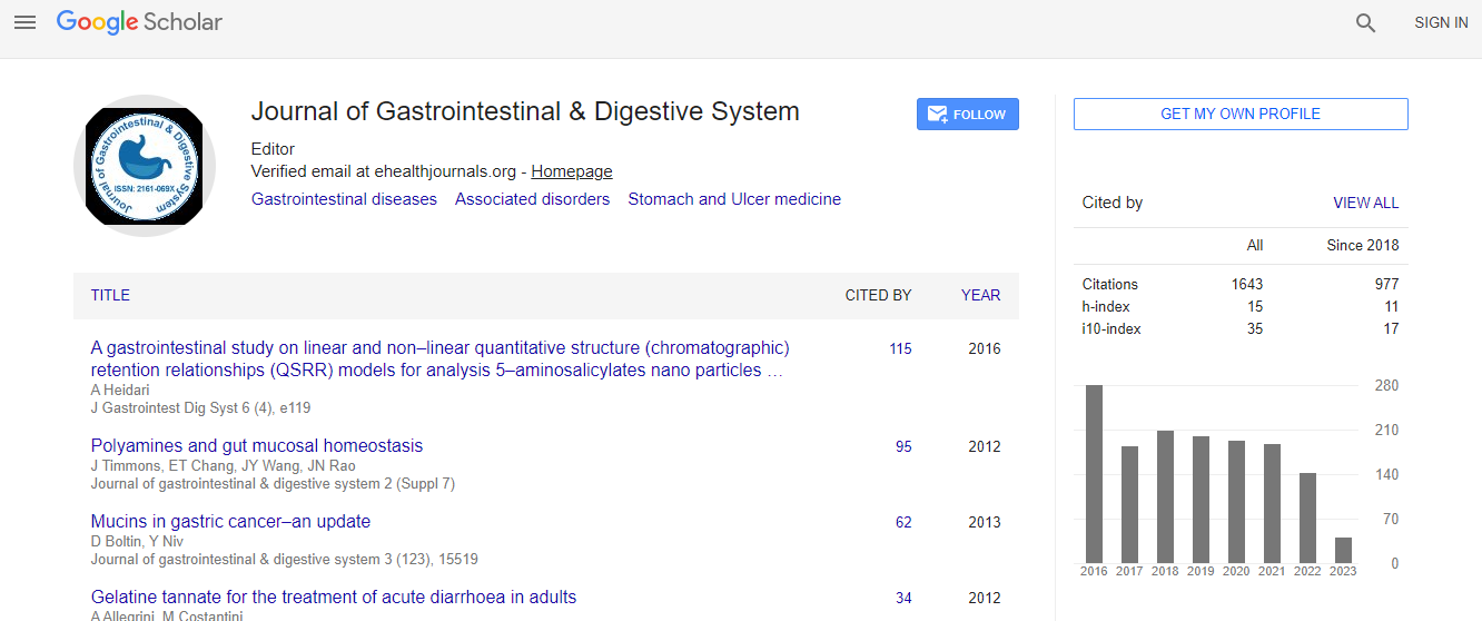
Google Scholar citation report
Citations : 2091
Journal of Gastrointestinal & Digestive System received 2091 citations as per Google Scholar report
Journal of Gastrointestinal & Digestive System peer review process verified at publons

Indexed In
- Index Copernicus
- Google Scholar
- Sherpa Romeo
- Open J Gate
- Genamics JournalSeek
- China National Knowledge Infrastructure (CNKI)
- Electronic Journals Library
- RefSeek
- Hamdard University
- EBSCO A-Z
- OCLC- WorldCat
- SWB online catalog
- Virtual Library of Biology (vifabio)
- Publons
- Geneva Foundation for Medical Education and Research
- Euro Pub
- ICMJE
Useful Links
Recommended Journals
Related Subjects
Share This Page
Quantitative analysis of microcirculation changes of hepatic ischemia reperfusion injury in rabbits with liver cirrhosis by contrast-enhanced ultrasound
Joint Event on 13th International Conference on Pediatric Gastroenterology Hepatology & Nutrition & 3rd International Conference on Digestive and Metabolic Diseases
Haiyuan Li
Medical Park Private Tarsus Hospital, Turkey
Posters & Accepted Abstracts: J Gastrointest Dig Syst
Abstract
Objective: To investigate hepatic microcirculation perfusion before and after hepatic ischemia reperfusion injury (IRI) in rabbits with liver cirrhosis by the quantitative analysis of contrast-enhanced ultrasound (CEUS). Methods: Forty-five New Zealand rabbits with liver cirrhosis were randomly divided into sham operation group (group A) and ischemia reperfusion injury group (group B and group C). CEUS examination, aspartate aminotransferase (AST), alanine aminotransferase (ALT) and histopathological examination were performed before and after reperfusion of 0h, 1h, 6h, 24h and 48h, respectively. SonoLiver software was used to perform the quantitative analysis of CEUS. And the time intensity curve (TIC) was used to measure peak intensity (IMAX), rise time (RT), peak time (TTP), respectively. Results: There were no significant differences in TIC parameters (IMAX, RT and TTP) in the group A at each time point (P > 0.05). IMAX in groups B and C at 0h, 1h, 6h, 24h, 48h of reperfusion had no significant change compared with the blocking (P > 0.05). In groups B and C, RT, TTP increased significantly at 0h, 1h, 6h of reperfusion compared with before reperfusion (P < 0.05), however, the parameters of RT and TTP had no changes at 24h of reperfusion had no obvious change than before reperfusion ,no significant difference (P > 0.05),48h of reperfusion compared with before reperfusion decreased, with statistical difference (P < 0.05). Pearson correlation analysis showed that RT and TTP were positively correlated with ALT and AST (P < 0.001). In groups B and C, the pathological changes at 0h of reperfusion showed edema of liver cells, liver sinusoidal space narrowing, gathered a large number of red blood cells in liver sinusoids and central vein blocking lumen, micro thrombosis. With the prolongation of reperfusion, the pathology revealed that red blood cell aggregation in hepatic sinusoids and the portal area, caused by occlusion of the lumen of the portal area, microcirculation, there is a small amount of neutrophils at 1h of reperfusion. The pathological analysis revealed that edema was found in liver cells increases in ballooning, and saw extensive infiltration of neutrophils when at 6h of reperfusion. Hepatocyte atrophy, necrosis and hepatic sinus collapse were found at 24h of reperfusion. Conclusion: The quantitative analysis of contrast-enhanced ultrasound is a noninvasive, objective and accurate method to evaluate the changes of hepatic IRI microcirculation.Biography
E-mail: yanghonggx@163.com