 Spanish
  Chinese
  Russian
  German
  French
  Japanese
  Portuguese
  Hindi
Hindi Our Group organises 3000+ Global Conferenceseries Events every year across USA, Europe & Asia with support from 1000 more scientific Societies and Publishes 700+ Open Access Journals which contains over 50000 eminent personalities, reputed scientists as editorial board members.
Open Access Journals gaining more Readers and Citations
700 Journals and 15,000,000 Readers Each Journal is getting 25,000+ Readers
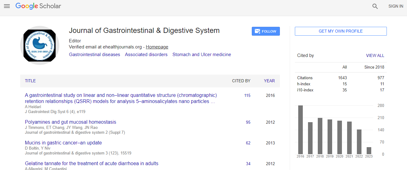
Google Scholar citation report
Citations : 2091
Journal of Gastrointestinal & Digestive System received 2091 citations as per Google Scholar report
Journal of Gastrointestinal & Digestive System peer review process verified at publons

Indexed In
- Index Copernicus
- Google Scholar
- Sherpa Romeo
- Open J Gate
- Genamics JournalSeek
- China National Knowledge Infrastructure (CNKI)
- Electronic Journals Library
- RefSeek
- Hamdard University
- EBSCO A-Z
- OCLC- WorldCat
- SWB online catalog
- Virtual Library of Biology (vifabio)
- Publons
- Geneva Foundation for Medical Education and Research
- Euro Pub
- ICMJE
Useful Links
Recommended Journals
Related Subjects
Share This Page
Physical tissue expansion for nanoscale investigation of clinical specimens
International Conference on Gastrointestinal Cancer and Therapeutics & 4th World Congress on Digestive & Metabolic Diseases & 26th Annual Congress on Cancer Science and Targeted Therapies
Octavian Bucur
BIDMC, USA
Keynote: J Gastrointest Dig Syst
Abstract
Objective: In pathology, examination of cellular structures and molecular composition using diffraction-limited microscopy is key to diagnosis. Recently, a new approach, Expansion Microscopy, was developed, enabling physical magnification and high resolution imaging of cell lines and mouse brain sections with conventional optical microscopes, by embedding them in a dense swellable polymer and adding water to swell the polymer after the enzymatic digestion of the proteins (Chen F et al., Science, 2015). The purpose of our study is to develop a pathology-optimized physical tissue expansion method for nanometer imaging and investigation of clinical tissue samples and to analyze its utility in diagnostic pathology and research. Methods: We developed a pathology optimized physical tissue expansion methods called Expansion Pathology (ExPath), which uses clinically optimized chemistry, labeling and imaging methodologies to expand and visualize both human FFPE and frozen clinical samples, including previously stained/unstained, mounted/unmounted and whole tissue slide/tissue microarrays sections, of a wide variety of fixed human tissue types and pathologies. Results: This ExPath protocol enabled expansion of human normal and cancer tissues (including gastrointestinal malignancies) ~4.5x in linear dimension and ~100x in volume, with a post-expansion measurement error of 3-7%. Physical tissue expansion pushes the optical microscopes beyond their limits (currently 250nm in resolution), by enabling for the first time ~70nm resolution imaging of diverse biomolecules in intact tissue with an optical microscope. With ExPath, certain lesions and pathologies previously diagnosed with an electron microscopy (EM) can now be diagnosed with a conventional optical microscope after physical tissue expansion, an inexpensive, faster and reliable strategy. It also enables high-fidelity computational discrimination between early neoplastic lesions that to date have challenged the human judgment. Conclusion: ExPath offers new approaches for assessing pathologically important features in human tissue. It may eliminate the need for EM in the diagnosis of certain diseases for which EM is required for diagnosis and it can improve the computational discrimination between pathological lesions that are hard to distinguish with existing techniques. ExPath may enable routine use of nanoscale imaging in molecular pathology and research (2017, Nature Biotechnology).Biography
Octavian Bucur, MD, PhD is Instructor in the Department of Pathology at the Harvard Medical School and Beth Israel Deaconess Medical Center, in Boston, MA, focusing on the development and application of new experimental and computational technologies with significant impact in molecular, diagnostic pathology and personalized medicine. He is also a member of the Ludwig Cancer Center at Harvard and Broad Institute of MIT and Harvard. In collaboration with Dr Edward Boyden’s laboratory at MIT, he has developed a pathology-optimized physical tissue expansion method called Expansion Pathology, that enables ~100 times expansion in volume of any type of clinical specimen and visualization of 70-80nm structures with conventional optical microscopes (currently limited to ~250nm resolution). Expansion Pathology has the potential of replacing electron microscopy in diagnosis and investigation of certain pathologies and nanometer structures (Nature Biotechnology, August 2017, 3 patents filed).
E-mail: obucur@bidmc.harvard.edu