Our Group organises 3000+ Global Conferenceseries Events every year across USA, Europe & Asia with support from 1000 more scientific Societies and Publishes 700+ Open Access Journals which contains over 50000 eminent personalities, reputed scientists as editorial board members.
Open Access Journals gaining more Readers and Citations
700 Journals and 15,000,000 Readers Each Journal is getting 25,000+ Readers
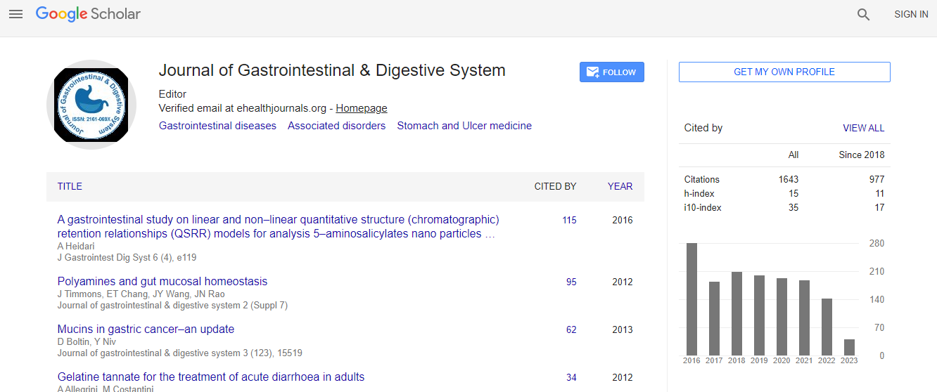
Google Scholar citation report
Citations : 2091
Journal of Gastrointestinal & Digestive System received 2091 citations as per Google Scholar report
Journal of Gastrointestinal & Digestive System peer review process verified at publons
Indexed In
- Index Copernicus
- Google Scholar
- Sherpa Romeo
- Open J Gate
- Genamics JournalSeek
- China National Knowledge Infrastructure (CNKI)
- Electronic Journals Library
- RefSeek
- Hamdard University
- EBSCO A-Z
- OCLC- WorldCat
- SWB online catalog
- Virtual Library of Biology (vifabio)
- Publons
- Geneva Foundation for Medical Education and Research
- Euro Pub
- ICMJE
Useful Links
Recommended Journals
Related Subjects
Share This Page
Observation of the pharynx to the cervical esophagus using transnasal endoscopy with image enhanced endoscopy
11th Global Gastroenterologists Meeting
Kenro Kawada
Tokyo Medical and Dental University, Japan
ScientificTracks Abstracts: J Gastrointest Dig Syst
Abstract
The more progress achieved in endoscopy, the more superficial cancers in the head and neck regions associated with esophageal squamous cell carcinoma have been found. Between August 1996 and March 2017, we have been experienced 350 cases of superficial head and neck cancers. Some areas difficult to observe with trans-oral endoscopy because of gag reflex. We applied new trans-nasal esophagogastroduodenoscopy (EGD) with image enhanced endoscopy (narrow band imaging, blue laser imaging, and linked color imaging) and modifications of endoscopic techniques for observing head and neck cancers and obtaining excellent results. The patient was asked to bow their head deeply in the left lateral position, and then we kept our hand on the back of the patient��?s head and pushed it forward by one span of our hand. Then, he was asked to lift up their chin as far as possible. After the local anesthesia of the nose without sedation, the endoscope was inserted through the nose. When inspecting the hypopharynx and the orifice of the esophagus, we asked the patient to blow hard and puff his cheeks with his mouth closed. This procedure provided a much better view of the orifice of the esophagus than had been possible with trans-oral endoscopy. Furthermore, observing the base of the tongue using trans-oral endoscopy is also difficult. When inspecting the oropharynx, the patient opens their mouth wide and sticks their tongue out as far as possible while making a vocal sound similar to a long I. The endoscopist then forces the transnasalendoscope to make a U-turn, and observes the oropharynx, in particular the base of the tongue. Mucosal redness, a pale thickened mucosa, white deposits, or loss of a normal vascular pattern, as well as demarcated brownish areas with image enhanced endoscopy, are important characteristics to diagnose superficial carcinoma.Biography
Kenro Kawada completed his Graduation in 1995 at Tokyo Medical and Dental University. He worked at Medical Hospital, Tokyo Medical and Dental University in 1995 and 2001. He was Junior Associate Professor in Department of Gastrointestinal Surgery at Tokyo Medical and Dental University in 2008.
Email: kawada.srg1@tmd.ac.jp

 Spanish
  Chinese
  Russian
  German
  French
  Japanese
  Portuguese
  Hindi
Hindi 
