 Spanish
  Chinese
  Russian
  German
  French
  Japanese
  Portuguese
  Hindi
Hindi Our Group organises 3000+ Global Conferenceseries Events every year across USA, Europe & Asia with support from 1000 more scientific Societies and Publishes 700+ Open Access Journals which contains over 50000 eminent personalities, reputed scientists as editorial board members.
Open Access Journals gaining more Readers and Citations
700 Journals and 15,000,000 Readers Each Journal is getting 25,000+ Readers
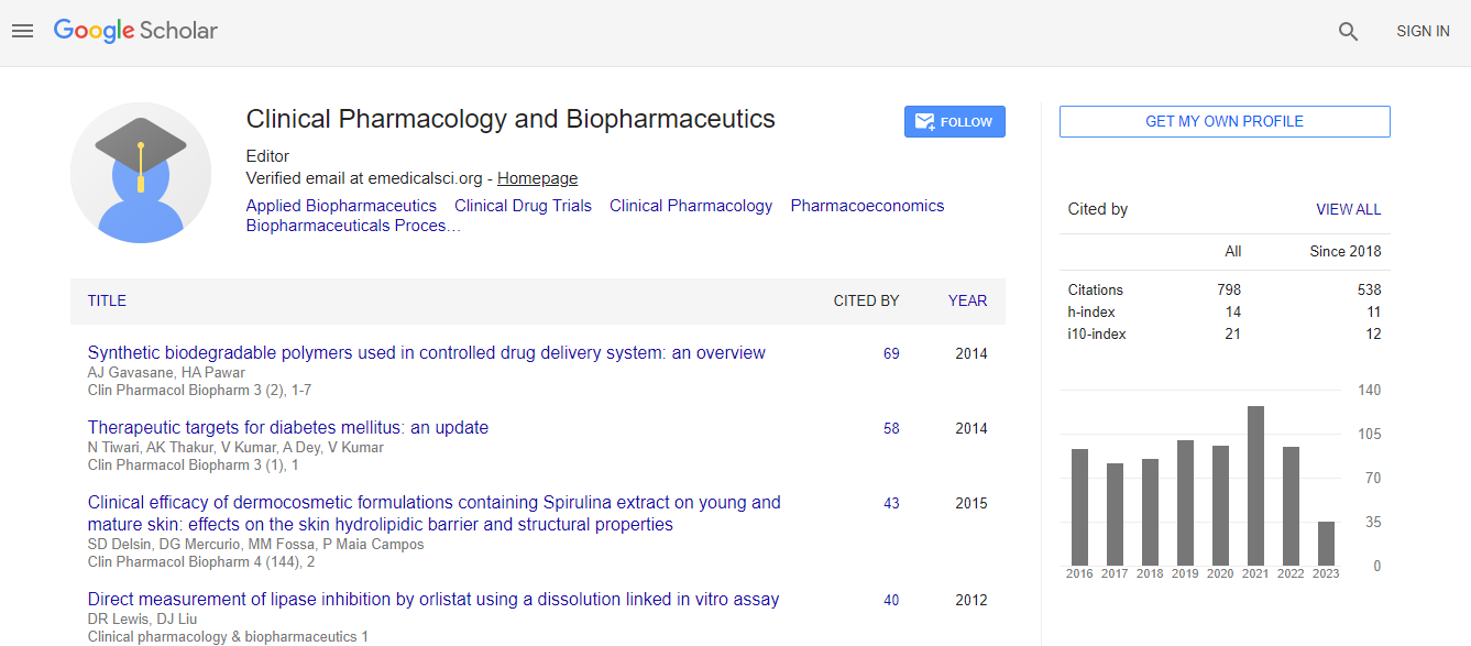
Google Scholar citation report
Citations : 1089
Clinical Pharmacology & Biopharmaceutics received 1089 citations as per Google Scholar report
Clinical Pharmacology & Biopharmaceutics peer review process verified at publons

Indexed In
- CAS Source Index (CASSI)
- Index Copernicus
- Google Scholar
- Sherpa Romeo
- Genamics JournalSeek
- RefSeek
- Hamdard University
- EBSCO A-Z
- OCLC- WorldCat
- Publons
- Euro Pub
- ICMJE
Useful Links
Recommended Journals
Related Subjects
Share This Page
Nanoparticles as alternate strategies for drug delivery into the Alzheimer brain: Electron microscopy qualitative ultrastructural analysis
International Conference and Expo on Biopharmaceutics
Gjumrakch Aliev
The University of Atlanta, USA
Posters-Accepted Abstracts: Clin Pharmacol Biopharm
Abstract
One of the biggest problems and challenges in the development of new drugs and/or treatment strategies for Alzheimer disease (AD) is the difficulty of passing the drugs across the blood brain barrier (BBB). The use of nanoparticles in drug delivery therapy holds much promise in targeting remote tissues, and as a result many studies have attempted to study the ultrastructural localization of nanoparticles in various tissues. However, there are currently no in vivo studies demonstrating the ultrastructural distribution of nanoparticles in the brain even in normal physiological conditions. The object of this study is to illuminate how injection of silver nanoparticles in the brain lead to leaking on the interendothelial contact and endothelial luminal plasma membrane, elucidating the possibility of penetrating into the areas of the brain which are most affected by AD (hippocampal tissues cellular compartment: vascular endothelium, perivascular, neuronal and glial cells). We investigated the ultrastructural distribution of nanoparticles [silver ion (5 nm), final concentration 0.9% which is dissolved in 0.9% NaCl,) in the rat brain hippocampal tissue one and four days after intra-peritoneal (i.p.) injection. The control experiment animals were given the same amount of vehicle treatment (0.9% NaCl, total amount 1 ml) for one and four days. At the end of the experiment perfusion fixation brain tissue was collected for electron microscopic analysis. Randomly selected ultrathin sections of the CA1 and CA3 areas with and without count staining were examined using a transmission electron microscope operating system at 100kV. Images were randomly selected and captured using a Digital Image system. Control animals which received vehicle injection revealed typical ultrastructural morphology of brain microvessels and neurons. The animals that received nanoparticles injection showed that after one and four days of silver injection, varying sizes of silver aggregates were seen throughout the neuronal cell bodies. Very often neuronal cell bodies were characterized by the absence of organelles, including mitochondria which coexisted with the presence of silver particles. The accumulation of the silver particles was also associated with the extracellular matrix, which was observed to coexist in the presence of a flake-like structure surrounding the neuronal tissue after four days of silver injection, and appeared to be a permanent feature of the hippocampal tissue. An interesting characteristic of the brain tissue appeared to be the disruption of the interendothelial contacts in an area where silver ions were present, which indicated that using nanoparticles in in vivo conditions is clearly undesirable in treatment, and that expanding this study in the future to include multiple dosages for comparison is necessary. Therefore this study can be basis for future conjugated silver nanoparticles drugs which able to deliver into the cell cytoplasm, therefore being able to diminish the lesions that occur in AD brain.Biography
Email: aliev03@gmail.com