Our Group organises 3000+ Global Conferenceseries Events every year across USA, Europe & Asia with support from 1000 more scientific Societies and Publishes 700+ Open Access Journals which contains over 50000 eminent personalities, reputed scientists as editorial board members.
Open Access Journals gaining more Readers and Citations
700 Journals and 15,000,000 Readers Each Journal is getting 25,000+ Readers
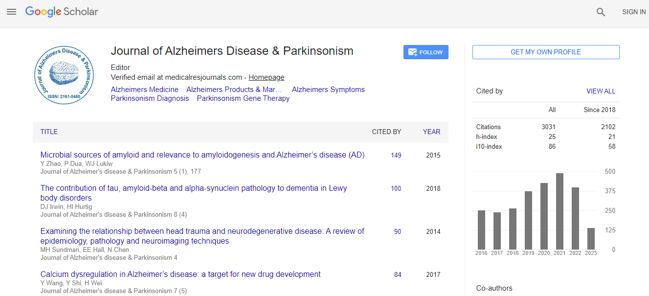
Google Scholar citation report
Citations : 4334
Journal of Alzheimers Disease & Parkinsonism received 4334 citations as per Google Scholar report
Journal of Alzheimers Disease & Parkinsonism peer review process verified at publons
Indexed In
- Index Copernicus
- Google Scholar
- Sherpa Romeo
- Open J Gate
- Genamics JournalSeek
- Academic Keys
- JournalTOCs
- China National Knowledge Infrastructure (CNKI)
- Electronic Journals Library
- RefSeek
- Hamdard University
- EBSCO A-Z
- OCLC- WorldCat
- SWB online catalog
- Virtual Library of Biology (vifabio)
- Publons
- Geneva Foundation for Medical Education and Research
- Euro Pub
- ICMJE
Useful Links
Recommended Journals
Related Subjects
Share This Page
MRI in dementia: Characteristics and relationship of atrophy and cognitive function
11th International Conference on Alzheimers Disease & Dementia
Nedim Ongun
Burdur State Hospital, Turkey
ScientificTracks Abstracts: J Alzheimers Dis Parkinsonism
Abstract
Introduction: The prevalence of dementia is rapidly increasing in developed countries because of significant increase in aging population. In order to be able to make a faster and more accurate decision, the importance of imaging in diagnosis and treatment is increasing steadily. Magnetic resonance imaging (MRI) is one method by which the extent, impact and possible etiology of regional brain pathology can be quantitatively assessed. The aim of this study is to reveal the imaging properties of dementia and investigate the relationship between MRI and cognitive score within the vascular dementia (VaD) and Alzheimer├ó┬?┬?s disease (AD). Methods: 1024 patients diagnosed with dementia were evaluated retrospectively and grouped according to clinical features. Demographic characteristics and risk factors were recorded. MRI scans were scored using Scheltens' scale and visual analogue scale. The Mini-Mental State Examination (MMSE) was used to assess the cognitive status. Patients were compared based on MRI measurements and MMSE scores. Results: 242 patients without an MRI were excluded. 398 patients were probably suffering from AD with NINCDS-ADRDA criteria; 249 patients were probably suffering from VaD with NINDS-AIREN criteria and 135 patients were with other dementia subgroups. In MRI ratings, global atrophy (GA) and white matter hyperintensity (WMH) scores were significantly higher in VaD group (p<0.001) and medial temporal atrophy (MTA) scores were significantly higher in AD group (p<0.001). Age, WMH and MTA were significantly related to GA in VaD and AD groups. While WMH and MTA were associated with both groups, age was also associated with MTA in AD group. MMSE scores were associated with MTA in both VaD and AD. There was a significant association between MMSE and WMH in the VaD group but not in the AD group. Conclusion: This study is important for the evaluation of dementia related imaging properties. We conclude that clinical dementia is accompanied by brain tissue loss, regardless of etiology. These results may affect future investigations aimed at primary or secondary prevention of VaD and AD. Although selected treatments may vary, clinical outcomes are likely to be tied to slowing or preventing brain tissue loss.Biography
Nedim Ongun has completed his Neurology Residency and is working as a Neurologist. He is also pursuing his PhD in Physiology at the university. He has published more than 25 papers in reputed journals and has been serving as a Referee and Editorial Board Member.
Email:nedimongun@yahoo.com

 Spanish
  Chinese
  Russian
  German
  French
  Japanese
  Portuguese
  Hindi
Hindi 
