Our Group organises 3000+ Global Conferenceseries Events every year across USA, Europe & Asia with support from 1000 more scientific Societies and Publishes 700+ Open Access Journals which contains over 50000 eminent personalities, reputed scientists as editorial board members.
Open Access Journals gaining more Readers and Citations
700 Journals and 15,000,000 Readers Each Journal is getting 25,000+ Readers
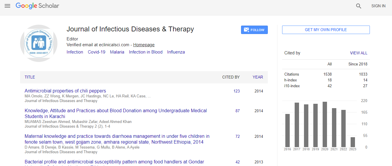
Google Scholar citation report
Citations : 1529
Journal of Infectious Diseases & Therapy received 1529 citations as per Google Scholar report
Indexed In
- Index Copernicus
- Google Scholar
- Open J Gate
- RefSeek
- Hamdard University
- EBSCO A-Z
- OCLC- WorldCat
- Publons
- Euro Pub
- ICMJE
Useful Links
Recommended Journals
Related Subjects
Share This Page
Meningitis, intracranial abscess and suppurative thrombophlebitis of the lateral and/or cavernous sinuses are dreadful complications of chronic infectious/inflammatory conditions of the middle ear: A rare case of meningitis caused by recurrent cholesteatoma
Joint Event on 2nd World Congress on Infectious Diseases & International Conference on Pediatric Care & Pediatric Infectious Diseases
Veeraraghavan Meyyur Aravamudan
National University Hospital, Singapore
Posters & Accepted Abstracts: J Infect Dis Ther
Abstract
A 46 year old Malay male with past surgical history of mastoidectomy in 2007 for cholesteatoma was admitted with sudden onset of headache, altered level of consciousness and lethargy for 1 day. Associated symptoms included multiple episodes of non-projectile vomiting and photophobia. He denied blurring of vision, otalgia and otorrhea. Physical examination revealed a lethargic looking male patient with a GCS of 3. His temperature was 38.5oC. Neck rigidity was present on movement in all directions. Cranial nerve and fundoscopic examination was unremarkable. He had skew deviation of eyes to right. Rest of the neurological examination did not reveal any motor deficits. He was started on empirical intravenous ceftriaxone, vancomycin, acyclovir and ampicillin for clinically suspected meningitis while awaiting lumbar puncture results. Computerized tomography (CT) of brain was normal. Cerebrospinal fluid (CSF) obtained from lumbar puncture showed cell count of 900 units per mm3 with 80% lymphocytes and 20% neutrophils, protein of 0.92 gram per liter and glucose was 1.7 mmol per liter. Gram stain did not reveal any organism and cultures were negative. Polymerase chain reaction (PCR) for neurotropic viruses i.e., HSV, measles, mumps and enterovirus were negative. Cerebrospinal fluid acid fast bacillus (AFB) smear was negative. Computerized tomography (CT) temporal bone with contrast showed right middle ear and mastoid cholesteatoma with surrounding infected granulation tissue which extended into the inner ear and intracranially. He was subsequently referred to ENT and underwent right modified radical mastoidectomy. He made a good clinical recovery. Cholesteatoma is a destructive and expanding growth consisting of keratinizing squamous epithelium in the middle ear and/or mastoid process. Because of their erosive and expansile properties they can destroy the ossicles and can potentially spread into the base of the skull into the brain causing meningitis. Even though the incidence of cholesteatoma causing meningitis is rare, these are still potential life threatening complications. Cholesteatoma is still considered a surgical disease requiring either the complete removal of its squamous lined matrix or its exteriorization for continued aural toilet and ventilation. In the pre-antibiotic era, the mortality rate from intracranial complications following the otologic diseases was approximately 75%. In the post-antibiotic era, mortality was around 34%. Meningitis is the most common intracranial complication. There are three dissemination passages for the occurrence of otogenic meningitis which are hematogenous, congenital dehiscence (such as Hyrtl├ó┬?┬?s fissures) or preformed (osseous erosion). Every patient with suspicion of complication needs to be followed up by several medical specialties and must be submitted to full physical exam and computerized tomography with contrast. The treatment must be aggressive with early initiation of intravenous antibiotic and early drainage of the infectious focus in order to reduce the morbidity and mortality rate. Early recognition and computerized tomography of temporal bone were important in diagnosis of meningitis secondary to cholesteatoma and prompt referral to ENT surgeon for early surgery should be considered to avoid long-term complications.Biography
Veeraraghavan Meyyur Aravamudan is senior resident in advanced internal medicine at National University Hospital, Singapore.
Email: drveerupaed2000@yahoo.co.in

 Spanish
  Chinese
  Russian
  German
  French
  Japanese
  Portuguese
  Hindi
Hindi 