ISSN: 2161-0681

Journal of Clinical & Experimental Pathology
Open Access

Our Group organises 3000+ Global Conferenceseries Events every year across USA, Europe & Asia with support from 1000 more scientific Societies and Publishes 700+ Open Access Journals which contains over 50000 eminent personalities, reputed scientists as editorial board members.

Open Access Journals gaining more Readers and Citations
700 Journals and 15,000,000 Readers Each Journal is getting 25,000+ Readers

This Readership is 10 times more when compared to other Subscription Journals (Source: Google Analytics)
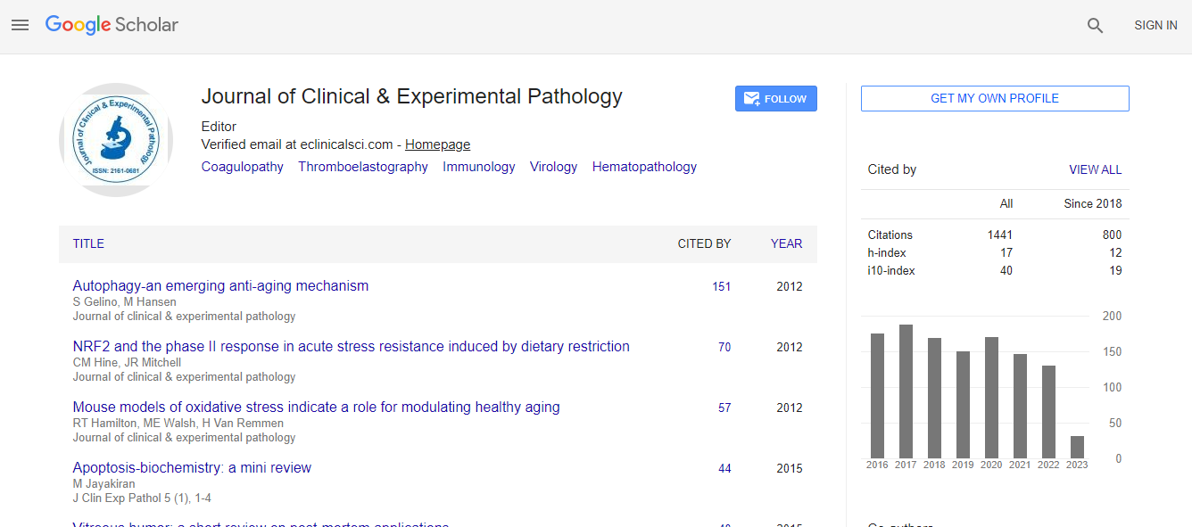
Google Scholar citation report
Citations : 1437

Journal of Clinical & Experimental Pathology received 1437 citations as per Google Scholar report

Journal of Clinical & Experimental Pathology peer review process verified at publons
Indexed In
  • Index Copernicus
  • Google Scholar
  • Sherpa Romeo
  • Open J Gate
  • Genamics JournalSeek
  • JournalTOCs
  • Cosmos IF
  • Ulrich's Periodicals Directory
  • RefSeek
  • Directory of Research Journal Indexing (DRJI)
  • Hamdard University
  • EBSCO A-Z
  • OCLC- WorldCat
  • Publons
  • Geneva Foundation for Medical Education and Research
  • Euro Pub
  • ICMJE
  • world cat
  • journal seek genamics
  • j-gate
  • esji (eurasian scientific journal index)
Share This Page

Juvenile ossifying fibroma of the maxilla: Differential diagnosis against well-differentiated osteosarcoma

4th International Conference and Exhibition on Pathology

Hidetake Tachinami1, Kei Tomihara1, Hrioaki Tsuno1, 2 and Makoto Noguchi1

Posters-Accepted Abstracts: J Clin Exp Pathol

DOI: 10.4172/2161-0681.S1.017

Abstract
Background: Ossifying fibroma is a well-demarcated lesion composed of fibro-cellular tissue and mineralized material of vary appearances. Juvenile ossifying fibroma (JOF) is rare histological variant of ossifying fibroma. It may be sometimes difficult to distinguish JOF from other variety of fibro-osseous lesion. In this report, we present a case of maxillary JOF with some sarcomatoid characteristics suggested well-differentiated osteosarcoma. Methods: A twenty four-year-old man with a slowly enlarging cheek swelling of right maxilla with some nasal obstruction was referred to our hospital. Status also presented swelling of intraoral site of right maxilla and no paresthesia or eye symptom. Imaging studies showed a multilocular lesion that extended from posterior maxilla to maxillary sinus. Knife edged root absorption of right upper molar was observed; through peripheral bone resorption was not invasive. Results: Partial maxillectomy was performed, as biopsy specimen was suggested a benign fibro-osseous lesion. Postoperative histology indicated suspicious components of malignant transformation, mitosis or cell invasion to bone area, partially in most of cell-rich observed. Conclusion: Particularly in young generation, there might be some difficulties to distinguish between JOF and welldifferentiated osteosarcoma histologically. It is considered those clinical feathers are as crucial as histological finding in these cases.
Biography
Hidetake Tachinami has completed his D.D.S at the age of 24 years from Nippon Dental University and postdoctoral studies from Toyama University School of Medicine. His research interests include oral lichen planus and oral lichenoid lesion.
Top