Our Group organises 3000+ Global Conferenceseries Events every year across USA, Europe & Asia with support from 1000 more scientific Societies and Publishes 700+ Open Access Journals which contains over 50000 eminent personalities, reputed scientists as editorial board members.
Open Access Journals gaining more Readers and Citations
700 Journals and 15,000,000 Readers Each Journal is getting 25,000+ Readers
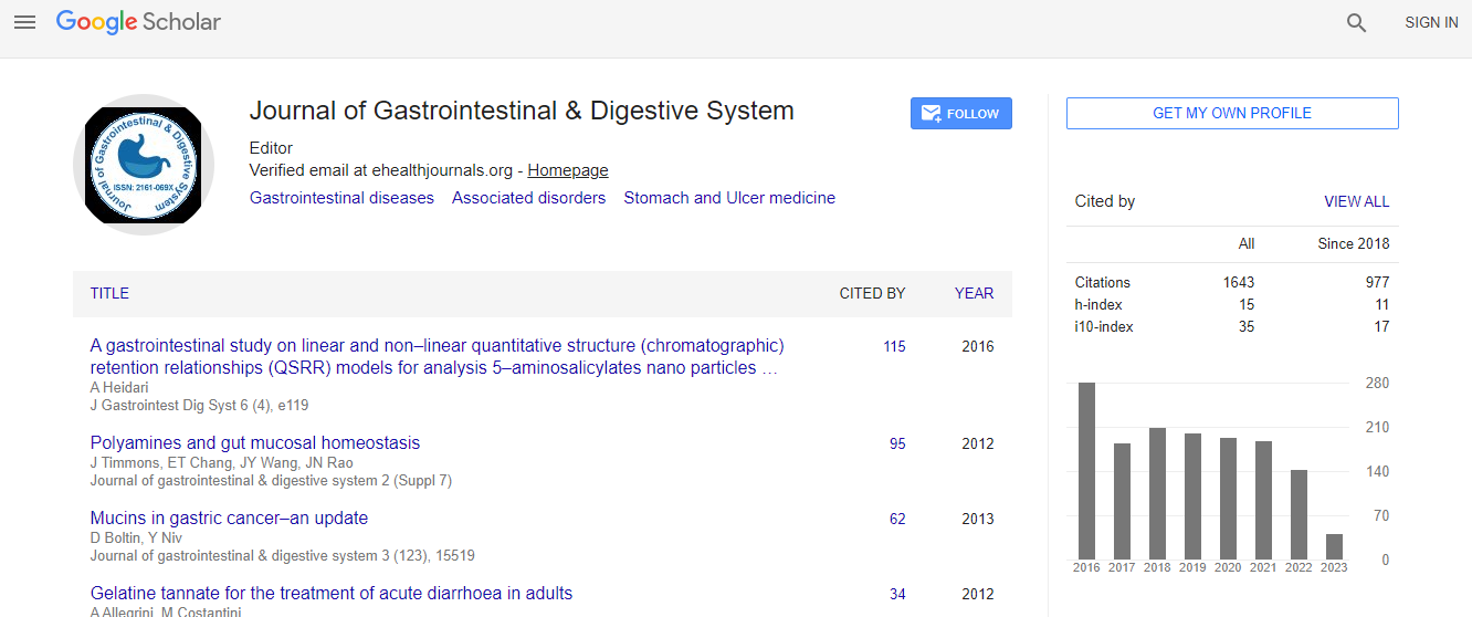
Google Scholar citation report
Citations : 2091
Journal of Gastrointestinal & Digestive System received 2091 citations as per Google Scholar report
Journal of Gastrointestinal & Digestive System peer review process verified at publons
Indexed In
- Index Copernicus
- Google Scholar
- Sherpa Romeo
- Open J Gate
- Genamics JournalSeek
- China National Knowledge Infrastructure (CNKI)
- Electronic Journals Library
- RefSeek
- Hamdard University
- EBSCO A-Z
- OCLC- WorldCat
- SWB online catalog
- Virtual Library of Biology (vifabio)
- Publons
- Geneva Foundation for Medical Education and Research
- Euro Pub
- ICMJE
Useful Links
Recommended Journals
Related Subjects
Share This Page
Imaging enhanced endoscopy: A practical approach
International Conference on Digestive Diseases
Lui Ka Luen
Tuen Mun Hospital, Hong Kong
ScientificTracks Abstracts: J Gastrointest Dig Syst
Abstract
Image enhanced endoscopy (IEE) is a combination of different advanced endoscopic methods which help to provide optical real time diagnosis for the luminal lesions. Traditional biopsy may have a disadvantage of sampling error since biopsy usually provide a small portion of lesion except the total excisional biopsy provided by small advanced excisional technique e.g, endoscopic submucosal dissection. Methods for IEE in general included special lighting e.g, narrow band imaging or blue laser imaging, both optical or digital magnification, chromoendoscopy and endoscopic ultrasound. Although different sites in luminal tract will have differences in terms of interpretion of IEE. In general, the purpose of IEE is to provide an optical diagnosis for a specific lesion regarding of the nature of lesion i.e., benign or malignant, and if the lesion is likely to have malignant component, the information about the depth of invasion can be provided. The approach of IEE start with the macroscopic appearance of the lesion in term of the color, shape, surface, consistency. The surface pattern and vascular pattern under the special light or chromoendoscopy are then observed to provide more information of the nature of the lesion and depth of invasion. Endoscopic ultrasound can provide more details of the depth of invasion. Good quality IEE is a must before any endoscopic treatment for the luminal lesion especially in the era of endoscopic submucosal dissection for good case selection.Biography
Lui Ka Luen completed his Graduation from University of Hong Kong in 2004 with distinction in Medicine. He became a Specialist in Gastroenterology in 2012 in Hong Kong and awarded Fellow of Hong Kong College of Physician in 2012. Then, he further pursued his career in “Imaging enhanced endoscopy, endoscopic ultrasound, endoscopic submucosal dissection and submucosal tunnel dissection” in Japan under direct mentorship of Professor Takashi Toyonaga. He is now an Honorary Clinical Assistant Professor at Chinese University of Hong Kong. He also published paper and is an invited speaker in various local and international journals, conferences and meetings.
Email: luikaluen@gmail.com

 Spanish
  Chinese
  Russian
  German
  French
  Japanese
  Portuguese
  Hindi
Hindi 
