Our Group organises 3000+ Global Conferenceseries Events every year across USA, Europe & Asia with support from 1000 more scientific Societies and Publishes 700+ Open Access Journals which contains over 50000 eminent personalities, reputed scientists as editorial board members.
Open Access Journals gaining more Readers and Citations
700 Journals and 15,000,000 Readers Each Journal is getting 25,000+ Readers
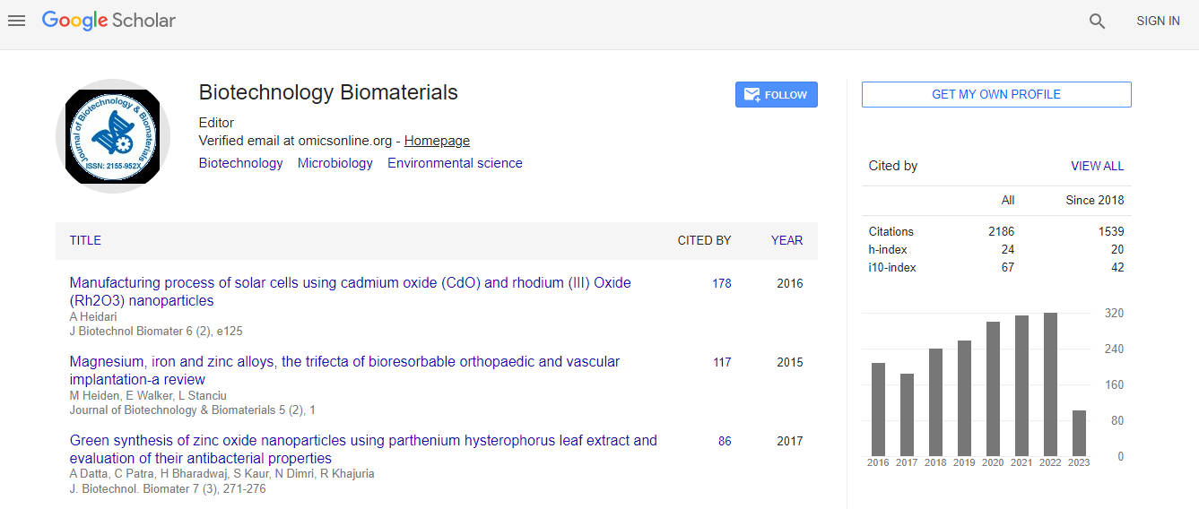
Google Scholar citation report
Citations : 3330
Journal of Biotechnology & Biomaterials received 3330 citations as per Google Scholar report
Indexed In
- Index Copernicus
- Google Scholar
- Sherpa Romeo
- Open J Gate
- Genamics JournalSeek
- Academic Keys
- ResearchBible
- China National Knowledge Infrastructure (CNKI)
- Access to Global Online Research in Agriculture (AGORA)
- Electronic Journals Library
- RefSeek
- Hamdard University
- EBSCO A-Z
- OCLC- WorldCat
- SWB online catalog
- Virtual Library of Biology (vifabio)
- Publons
- Geneva Foundation for Medical Education and Research
- Euro Pub
- ICMJE
Useful Links
Recommended Journals
Related Subjects
Share This Page
Free floating brain sections for immunofluorescence markers: A technical and scientific approach
17th EURO BIOTECHNOLOGY CONGRESS
Emmanuel Loeb
Patho-Logica, Scientific Park Ness Ziona, Israel
ScientificTracks Abstracts: J Biotechnol Biomater
Abstract
Free floating sections is regarded as a new histological method that can be used for immune fluorescence staining. This method is clearly the best way to go for optimal Ab expression in the tissue. Furthermore, staining of thick sections can later on be used for a confocal microscopical analysis. This presentation covers the technical work pattern of the method starting with the tissue preparation and conservation, threw brain accurate dissection and staining. The method is very suitable for morphometry quantification of histological data, here method of image analysis will be presented and the scientific value will be discussed. Furthermore, examples are presented of projects that had combined the method such as stroke and Parkinson models in lab animals. Finally a discussion will be presented were the advantages of the current method will be pointed compared to the classical immunohistochemistry methods.Biography
Emmanuel Loeb is a graduate from School of Veterinary Medicine, Utrecht University, Netherlands and a qualified expert, Veterinary Pathologist with published papers. He has 12 years of experience in Experimental Pathology and is constantly improving his skills through continuous profession development. In his work, he takes part in annual professional meetings such as the ESVP and follows The Society of Toxicologic Pathology recommendations. He established new methods in the laboratory such as “free floating sections” for immunofluorescence staining, and developed translation tools from pathological hallmarks to histological end point. He is also teaching pathology at the Veterinary School of Koret (Hebrew University).

 Spanish
  Chinese
  Russian
  German
  French
  Japanese
  Portuguese
  Hindi
Hindi 
