Our Group organises 3000+ Global Conferenceseries Events every year across USA, Europe & Asia with support from 1000 more scientific Societies and Publishes 700+ Open Access Journals which contains over 50000 eminent personalities, reputed scientists as editorial board members.
Open Access Journals gaining more Readers and Citations
700 Journals and 15,000,000 Readers Each Journal is getting 25,000+ Readers
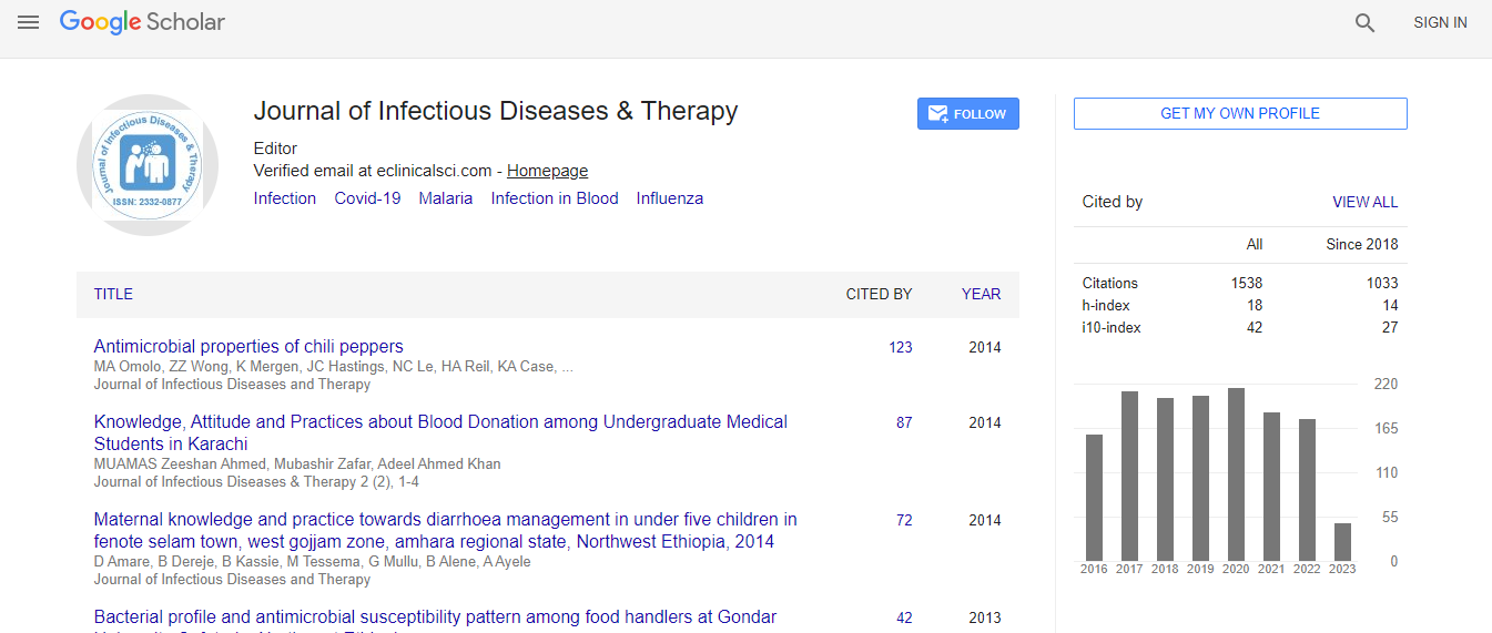
Google Scholar citation report
Citations : 1529
Journal of Infectious Diseases & Therapy received 1529 citations as per Google Scholar report
Indexed In
- Index Copernicus
- Google Scholar
- Open J Gate
- RefSeek
- Hamdard University
- EBSCO A-Z
- OCLC- WorldCat
- Publons
- Euro Pub
- ICMJE
Useful Links
Recommended Journals
Related Subjects
Share This Page
First isolation of Anaplasma phagocytophilum AAIK from Apodemus agrarius in Korea
World Congress on Infectious Diseases
Joon-Seok Chae1*, Sung-Suck Oh1, 2, Jeong-Byoung Chae1, Myeong-Je Hur2, Yun-Kyung Cho and Kyoung-Seong Choi3
ScientificTracks Abstracts: J Infect Dis Ther
Abstract
Human granulocytic anaplasmosis, an emerging infectious disease in the Republic of Korea (ROK), is caused by an obligate
intracellular tick-borne bacterium of the family Anaplasmataceae. Anaplasma phagocytophilum has been found in
a variety of animal species including wild deer, cats, dogs, gray squirrels, horses, and mice. The objective of the present study
was to isolate and characterize the strains of A. phagocytophilum in black-striped field mice (Apodemus agrarius) and Korean
water deer (Hydropotes inermis argyropus) in the ROK. Anaplasma infection based on the 16S rRNA genes were detected by
conventional polymerase chain reaction (PCR) and species-specific nested PCR assays with DNA from Korean water deer
(KWD) blood and black-striped field mice (BSFM) spleen. For isolation of A. phagocytophilum, BALB/c mice were used for
propagation. White blood cells from 4-5 infected BALB/c mice were inoculated into 3×10⁴ HL60 and THP-1 cell line, respectively.
Eight KWD and 27 BSFM were captured. Seven (87.5%) blood samples of KWD and 12 (44.4%) spleens of BSFM were
positive for A. phagocytophilum according to PCR. However, 7 white blood cell samples of KWD were not propagated in HL60
and THP-1 cell line. Twelve spleen suspensions of BSFM were pooled for inoculum, and 0.3 ml of spleen suspension was intraperitoneally
inoculated into 20 BALB/c mice (5 in each group; total 4 groups). When tested for 10 days post-inoculation,
inoculated mice were positive for A. phagocytophilum by PCR. Three of 4 groups were not propagated and 1 of 4 groups was
propagated in THP-1 cell line. A. phagocytophilum was observed in Wright-Giemsa stain preparations at 9 days after inoculation
of cell cultures. Morulae were found on 70% of THP-1 cells at 12 days post-inoculation. Cultured A. phagocytophilum were
confirmed by indirect immunofluorescence assay. PCR was performed using 16S rRNA, ankA, groEL, and msp2 gene primers
were used to amplify the genes of A. phagocytophilum. All of the nucleotide sequences from cultured isolate were identical
with A. phagocytophilum. We isolated the strain of A. phagocytophilum (AAIK isolate) in black-striped field mice (Apodemus
agrarius) in the ROK. This strain may be expected to contribute to public health and veterinary medicine.
Biography
Joon-seok Chae has completed his DVM, MS, PhD from Chonbuk National University and postdoctoral studies from Texas A&M University and University of
California-Davis. He is the Professor of College of Veterinary Medicine, Seoul National University. He has published more than 160 papers in reputed journals
and serving as an editorial board member of repute. His recent interesting research areas are tick-borne zoonotic pathogens (Anaplasma, Ehrlichia, Rickettsia,
Bartonella, TBE and SFTS virus etc.) and equine stem cell therapy.

 Spanish
  Chinese
  Russian
  German
  French
  Japanese
  Portuguese
  Hindi
Hindi 