Our Group organises 3000+ Global Conferenceseries Events every year across USA, Europe & Asia with support from 1000 more scientific Societies and Publishes 700+ Open Access Journals which contains over 50000 eminent personalities, reputed scientists as editorial board members.
Open Access Journals gaining more Readers and Citations
700 Journals and 15,000,000 Readers Each Journal is getting 25,000+ Readers
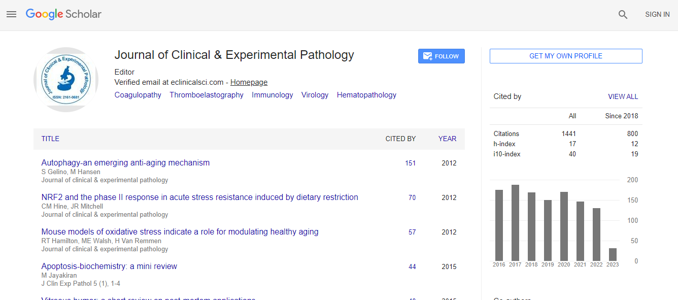
Google Scholar citation report
Citations : 2975
Journal of Clinical & Experimental Pathology received 2975 citations as per Google Scholar report
Journal of Clinical & Experimental Pathology peer review process verified at publons
Indexed In
- Index Copernicus
- Google Scholar
- Sherpa Romeo
- Open J Gate
- Genamics JournalSeek
- JournalTOCs
- Cosmos IF
- Ulrich's Periodicals Directory
- RefSeek
- Directory of Research Journal Indexing (DRJI)
- Hamdard University
- EBSCO A-Z
- OCLC- WorldCat
- Publons
- Geneva Foundation for Medical Education and Research
- Euro Pub
- ICMJE
- world cat
- journal seek genamics
- j-gate
- esji (eurasian scientific journal index)
Useful Links
Recommended Journals
Related Subjects
Share This Page
Electron Paramagnetic Resonance (EPR) spectroscopy for diagnosis and characterization of mitochondrial dysfunction and diseases
12th International Conference on Pediatric Pathology & Laboratory Medicine
Brian Bennett
Marquette University, USA
Keynote: J Clin Exp Pathol
Abstract
Mitochondrial disease (MD) presents with a wide range of clinical, pathological and biochemical outcomes and is consequently very difficult to diagnose conclusively. EPR is a magnetic resonance technique that detects and characterizes unpaired electrons that are present in transition metal ions in certain oxidation states {e.g. Fe(III), Cu(II) and Mn(II)}, clusters (e.g., [2Fe2S]+ red, [3Fe4S]+ ox, [4Fe4S]+ red) and free radicals (e.g., UQâ�?¢â�?�?, FADHâ�?¢). The mitochondrial respiratory chain complexes I-IV contain 23 potentially paramagnetic centers that exhibit distinct EPR signals depending on their redox potentials, the availability of electrons, the catalytic competence of each of the enzymatic complexes and the integrity of the electron transport chain (ETC). In addition, EPR signals may be observed from UQâ�?¢â�?�?, and from the [3Fe4S]+ cluster of m-aconitase that arises due to oxidative stress. Key factors thought to be involved in the symptoms and pathology of MD is lowered ATP production and the production of toxic reactive oxygen species (ROS). Either or both of these can occur when electron transfer is impeded due to lowered expression, lowered activity, or structural alteration of ETC complexes, or compromised ingress or egress of reducing equivalents. EPR of rapidly-frozen fresh biopsy tissue is uniquely able to provide a snapshot of the electron distribution among the redox centers in the functioning mitochondrial ETC against a background of other biochemical and pathological assays. We recently described the first application of this methodology to a rat model of MD and will here describe progress toward translation of the approach for diagnosis and differentiation of MDs in children.Biography
Brian Bennett has completed his BA and MA in Natural Sciences from the University of Cambridge and DPhil in Biochemistry from the University of Sussex. Currently, he is a Chair and Distinguished Professor of Physics at Marquette University, USA.
Email: brian.bennett@marquette.edu

 Spanish
  Chinese
  Russian
  German
  French
  Japanese
  Portuguese
  Hindi
Hindi 
