Our Group organises 3000+ Global Conferenceseries Events every year across USA, Europe & Asia with support from 1000 more scientific Societies and Publishes 700+ Open Access Journals which contains over 50000 eminent personalities, reputed scientists as editorial board members.
Open Access Journals gaining more Readers and Citations
700 Journals and 15,000,000 Readers Each Journal is getting 25,000+ Readers
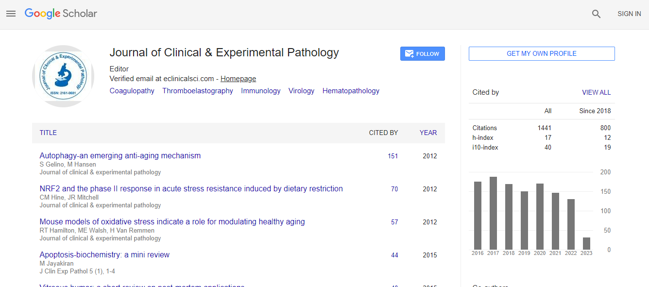
Google Scholar citation report
Citations : 2975
Journal of Clinical & Experimental Pathology received 2975 citations as per Google Scholar report
Journal of Clinical & Experimental Pathology peer review process verified at publons
Indexed In
- Index Copernicus
- Google Scholar
- Sherpa Romeo
- Open J Gate
- Genamics JournalSeek
- JournalTOCs
- Cosmos IF
- Ulrich's Periodicals Directory
- RefSeek
- Directory of Research Journal Indexing (DRJI)
- Hamdard University
- EBSCO A-Z
- OCLC- WorldCat
- Publons
- Geneva Foundation for Medical Education and Research
- Euro Pub
- ICMJE
- world cat
- journal seek genamics
- j-gate
- esji (eurasian scientific journal index)
Useful Links
Recommended Journals
Related Subjects
Share This Page
Digital pathology image analysis: Challenges and opportunities
6th European Pathology Congress
Nasir Rajpoot
University Hospitals Coventry & Warwickshire, UK
Posters & Accepted Abstracts: J Clin Exp Pathol
Abstract
The emerging discipline of digital pathology is poised to change the status quo in pathology practice for the better. A pathology department in a medium sized hospital deals with a workload of about 100,000 tissue slides every year, resulting in approximately 50 TB of high-resolution image data, compressed in a near lossless manner, per annum. The sheer size of multi-gigapixel images produced by digital slide scanners poses interesting technical challenges. On the other hand, the heap of image data linked with associated clinical and genomic data is a potential goldmine of invaluable information, as each high resolution image contains information about tens of thousands of cells and their spatial relationships with each other. I will present some of the recent developments in our group concerning digital pathology image analysis and tissue morphometrics from images of cancerous tissue slides. I will then discuss some of the main challenges in digital pathology and opportunities for exploring new unchartered territories.Biography
Email: N.M.Rajpoot@warwick.ac.uk

 Spanish
  Chinese
  Russian
  German
  French
  Japanese
  Portuguese
  Hindi
Hindi 
