Our Group organises 3000+ Global Conferenceseries Events every year across USA, Europe & Asia with support from 1000 more scientific Societies and Publishes 700+ Open Access Journals which contains over 50000 eminent personalities, reputed scientists as editorial board members.
Open Access Journals gaining more Readers and Citations
700 Journals and 15,000,000 Readers Each Journal is getting 25,000+ Readers
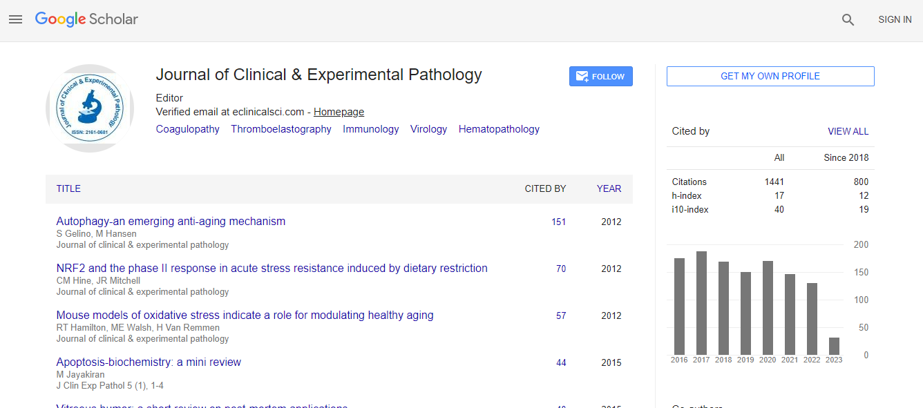
Google Scholar citation report
Citations : 2975
Journal of Clinical & Experimental Pathology received 2975 citations as per Google Scholar report
Journal of Clinical & Experimental Pathology peer review process verified at publons
Indexed In
- Index Copernicus
- Google Scholar
- Sherpa Romeo
- Open J Gate
- Genamics JournalSeek
- JournalTOCs
- Cosmos IF
- Ulrich's Periodicals Directory
- RefSeek
- Directory of Research Journal Indexing (DRJI)
- Hamdard University
- EBSCO A-Z
- OCLC- WorldCat
- Publons
- Geneva Foundation for Medical Education and Research
- Euro Pub
- ICMJE
- world cat
- journal seek genamics
- j-gate
- esji (eurasian scientific journal index)
Useful Links
Recommended Journals
Related Subjects
Share This Page
Cytomorphological spectrum of cysticercosis: A study of 72 cases
5th International Conference on Pathology
Anshoo Agarwal, Arvind Sinha and Amish Pawanarkar
RAK Medical and Health Sciences University, UAE
Posters & Accepted Abstracts: J Clin Exp Pathol
Abstract
Introduction: Cysticercosis is a worldwide infection caused by larval stage of a cestode, Taenia solium. Worm infestation is acquired by ingestion of undercooked pork containing the cysticerci. Cysticercosis is a common disease in most developing countries. It has its greatest prevalence in Mexico, other areas of Latin America, India, China, Africa and Europe. Cysticerci may present as single or multiple painless swellings in any organ or tissue of the body. The most common sites in order of frequency are the subcutaneous tissue, brain, muscle, heart, liver, lungs and peritoneum. These are usually mistaken clinically for dermatofibroma, neurofibroma, sebaceous cyst, dermoid cyst and calcified lymph nodes. Biopsy is a gold standard for definitive diagnosis of any lesion but nowadays fine needle aspiration cytology (FNAC) in the diagnosis of various parasitic lesions is well documented. In the present study we report clinical profile and cytomorphologic spectrum of cysticercosis findings on fine needle aspirates from 72 cases diagnosed as cysticercosis. Material & Methods: Over the period of 6 years, 72 cases of cysticercosis were diagnosed in the Department of Pathology, BPKIHS, Nepal & Shrivalli Nursing Home, Thane West, Maharashtra, India. All the patients presented with swellings of different regions of the body. FNA was performed with 22 gauge needle and 10 ml disposable plastic syringe. Aspirated materials were smeared onto the glass slides. Two slides were fixed immediately in 95% ethyl alcohol and stained with Papanicolaou stain. Two air dried smears were stained with May-Grunwald-Giemsa stain. Cases which were biopsied were processed for histopathological examination, stained with hematoxylin and eosin. Results: 38 patients were males and 34 patients were females. The age ranged from 1.5 to 76 years with majority of the patients (76.38%) being younger than 40 years of age. Most frequently affected site was upper extremity (47.28%). In 7 cases (9.73%) lingual cysticercosis was diagnosed in our study. Involvement of breast was seen in 4 cases (5.56%) which is a rare presentation. Clinically (98.7%) cases presented with a solitary lesion in the present study. Fine needle aspirates in our study yielded clear fluid in (32.27%) cases, blood mixed aspirate in (23.01%) cases and pus like aspirate in (44.72%) cases. Fragments are bluish fibrillary glial like structure. Outer wall layer was seen thrown into rounded wavy folds with tiny ovoid nuclei in a fribrillary stroma comprising of thin reticulin fibrils beneath it. In rest two of the cases (2.77%) diagnosis was suggested on associated other cytomorphologic features and inflammatory reaction comprising of eosinophils, neutrophils, histiocytes, epithelioid cells, lymphocytes and giant cells in varying proportions which were confirmed later on biopsy. In our study 24 cases (33.33%) were misdiagnosed clinically as cases of dermatofibroma, neurofibroma and sebaceous cyst. Conclusion: FNA cytology is a simple and reliable procedure for the diagnosis of cysticercosis. In principle, a mass produced by cestode should not be diagnosed by FNAB since it might cause anaphylaxis and/or dissemination of parasites.Biography
Email: dranshoo3@yahoo.com

 Spanish
  Chinese
  Russian
  German
  French
  Japanese
  Portuguese
  Hindi
Hindi 
