ISSN: 2161-0460

Journal of Alzheimers Disease & Parkinsonism
Open Access

Our Group organises 3000+ Global Conferenceseries Events every year across USA, Europe & Asia with support from 1000 more scientific Societies and Publishes 700+ Open Access Journals which contains over 50000 eminent personalities, reputed scientists as editorial board members.

Open Access Journals gaining more Readers and Citations
700 Journals and 15,000,000 Readers Each Journal is getting 25,000+ Readers
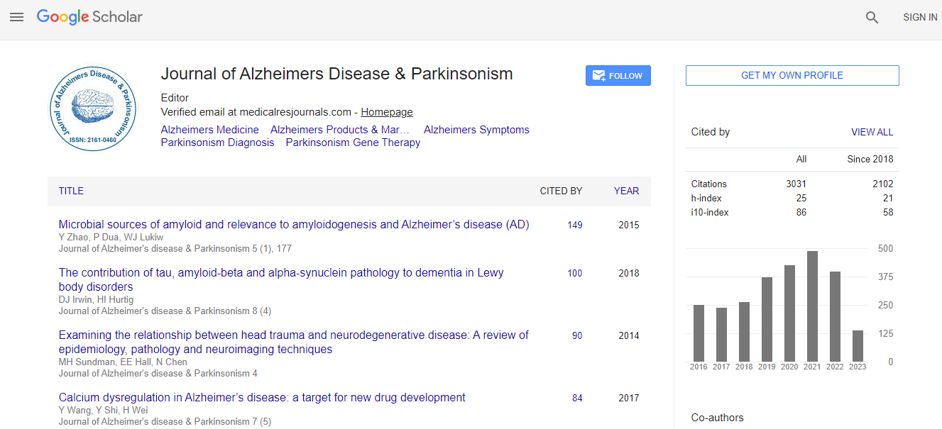
This Readership is 10 times more when compared to other Subscription Journals (Source: Google Analytics)

Cerebral blood flow by means of xenon-enhanced computed tomography: From trans-axial to surface quantitative images

2nd International Conference on Alzheimers Disease and Dementia

Shigeru Sase

ScientificTracks Abstracts: J Alzheimers Dis Parkinsonism

DOI: 10.4172/2161-0460.S1.006

Abstract
Each of the four major forms of dementia (Alzheimer�s disease, vascular dementia, dementia with Lewy body, and frontotemporal lobe degeneration) has been reported to have different pattern of cerebral blood flow (CBF), and could be distinguished from each other with the use of CBF images. Xenon-enhanced computed tomography (Xe-CT) can create quantitative CBF images like positron emission tomography (PET). Using newly developed volume CT, the whole brain CT image was obtained with one rotation (one scan). The whole brain CT image consisted of thin CT images. In the Xe-CT study, 30% non-radioactive xenon gas was inhaled for 4 minute, followed by 4-min breathing of air, in the meantime CT scan was conducted at 1 minute intervals. Corresponding to each individual CT image composing the whole CT image, CBF and partition coefficient (lambda) images were obtained. We developed a method of creating brain surface images of CT, CBF and lambda by piling thin layer images. With removing thin layers from the surface one by one, we could observe the changes in CT, CBF and lambda images from the surface to the inner area. By combining brain surface images with three kinds of tomographic images (trans-axial, sagittal, and coronal), and by combining neuronal activity information from CBF, foreign substance accumulation information from lambda, and morphological information from CT; we consider that it would be possible to establish a method of specifying the form of dementia and to detect the dementia in its early stage.
Biography
Shigeru Sase graduated from Tokyo Institute of Technology (Bachelor�s degree in 1975, and Master�s degree in 1977). He obtained PhD degree in medicine in 2002 at Toho University School of Medicine. He has been Guest Assistant Professor at the Faculty of Medicine, Toho University since 2004. He has been working for Anzai Medical Co., Ltd. since 1990. He has published many papers related to Xe-CT for the brain, liver and lung which appear in PubMed.
https://bahigox.fun/ https://bahisjet.fun/ https://bahsegel.fun/ https://betboo.fun/ https://betdoksan.xyz/ https://betebetgiris.xyz/ https://betexper.fun/ https://betgram.fun/ https://betkanyon.fun/ https://betkolik.fun/
Top