 Spanish
  Chinese
  Russian
  German
  French
  Japanese
  Portuguese
  Hindi
Hindi Our Group organises 3000+ Global Conferenceseries Events every year across USA, Europe & Asia with support from 1000 more scientific Societies and Publishes 700+ Open Access Journals which contains over 50000 eminent personalities, reputed scientists as editorial board members.
Open Access Journals gaining more Readers and Citations
700 Journals and 15,000,000 Readers Each Journal is getting 25,000+ Readers
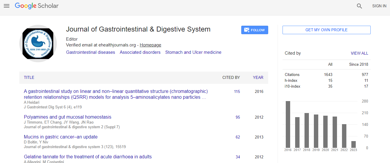
Google Scholar citation report
Citations : 2091
Journal of Gastrointestinal & Digestive System received 2091 citations as per Google Scholar report
Journal of Gastrointestinal & Digestive System peer review process verified at publons

Indexed In
- Index Copernicus
- Google Scholar
- Sherpa Romeo
- Open J Gate
- Genamics JournalSeek
- China National Knowledge Infrastructure (CNKI)
- Electronic Journals Library
- RefSeek
- Hamdard University
- EBSCO A-Z
- OCLC- WorldCat
- SWB online catalog
- Virtual Library of Biology (vifabio)
- Publons
- Geneva Foundation for Medical Education and Research
- Euro Pub
- ICMJE
Useful Links
Recommended Journals
Related Subjects
Share This Page
Calretinin Expression in Hirsch sprung Disease - A Potential Marker of Ganglion Cells
9th Euro Global Gastroenterology Conference
Mishal Sikandar
University of Health Sciences, Lahore, Pakistan
Posters & Accepted Abstracts: J Gastrointest Dig Syst
Abstract
Statement of problem: Hirschsprung disease (HSCR) or congenital intestinal aganglionosis is a birth defect characterized by complete absence of neuronal ganglion cells from a portion of the intestinal tract, mostly in a segment of rectum and variable length of contiguous proximal, causing functional obstruction and colonic dilation proximal to affected segment. (Amiel and Lyonnet, 2001) Routine diagnostic modalities like Haematoxylin & Eosin (H&E) and Acetylcholine Esterase (AChE) staining as well as radiology-based clinical techniques have been conventionally used for identification of aganglionosis and presence of hypertrophic nerve trunks in the affected segment as primary indicators of HSCR. (Kacar et al., 2012) However, these conventional methods have their inherent deficiencies as H&E requires multiple trans-mural biopsies and the interpretation of ganglion cells is often very difficult. (Tabbers et al., 2014) Similarly, AChE requires fresh frozen section for which the chances of technical error are very high and this facility is not commonly available in Pakistan. The number of misdiagnosed results with potential overtreatment stands in need for reliable staining to prevent harm from unnecessary surgery and mortality. Recently, Immunohistochemical markers are being increasingly used and evaluated in Pathology laboratories. No immunohistochemical marker, either alone or in combination, has emerged from those researches that are as promising as Calretinin. Hence, this study was designed with an aim to observe the immunohistochemical expression of Calretinin as a marker for aganglionosis and to detect ganglion cells in the affected areas for better and more accurate diagnosis of the disease. Methodology: This study was conducted at University of Health Sciences Lahore, Pakistan from February to September, 2016. Colonic Biopsy Specimens from 73 patients collected mostly from Mayo Hospital, Lahore with established histopathologic diagnosis of HSCR were considered for the study. Age range was 0.1-120months. There were 48(65.8%) cases who were â�?¤12 months old, 20(27.4%) were 12.1-60 months old and 5(6.8%) of the cases were 60.1-120 month old. The mean age of patients was 12.52�?±9.21 months. There were 52(71.23%) male and 21(28.77%) female patients. The male to female ratio in this study was 2.48:1. According to sign and symptoms and clinical examination, 69(94.5%) cases had mostly long standing constipation, 47(64.4%) cases had fever, 68(93.2%) cases had vomiting, 31(42.5%) cases had failure to thrive, 20(27.4%) cases had Enterocolitis and 63(86.3%) of the patients had palpable abdominal masses. Methodology involved staining of fresh sections with H&E procedure for provisional histological diagnosis. The biopsies were then processed for immunohistochemical staining with Calretinin and were observed for presence of ganglion cells. Findings: All the ganglion cells took brownish-black stain and were easily identified, which were not being identified on H&E. Ganglion cells were present and absent in 42(57.53%) and 31(42.47%) respectively. The study revealed that the Calretinin immunohistochemistry was very sensitive and specific for detecting ganglion cells. Conclusion and significance: It was concluded that Calretinin provides a very reliable and cost effective adjunctive test to be routinely used with H&E in the evaluation of Rectal Section Biopsies (RSBs) for HSCR. The use of Calretinin may help the Pathologists in making accurate and reliable diagnosis for HSCR and consequently eliminating the need for repeated biopsies and unnecessary surgeries.Biography
Email: mishal_sikandar@yahoo.com