Our Group organises 3000+ Global Conferenceseries Events every year across USA, Europe & Asia with support from 1000 more scientific Societies and Publishes 700+ Open Access Journals which contains over 50000 eminent personalities, reputed scientists as editorial board members.
Open Access Journals gaining more Readers and Citations
700 Journals and 15,000,000 Readers Each Journal is getting 25,000+ Readers
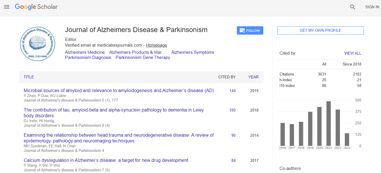
Google Scholar citation report
Citations : 4334
Journal of Alzheimers Disease & Parkinsonism received 4334 citations as per Google Scholar report
Journal of Alzheimers Disease & Parkinsonism peer review process verified at publons
Indexed In
- Index Copernicus
- Google Scholar
- Sherpa Romeo
- Open J Gate
- Genamics JournalSeek
- Academic Keys
- JournalTOCs
- China National Knowledge Infrastructure (CNKI)
- Electronic Journals Library
- RefSeek
- Hamdard University
- EBSCO A-Z
- OCLC- WorldCat
- SWB online catalog
- Virtual Library of Biology (vifabio)
- Publons
- Geneva Foundation for Medical Education and Research
- Euro Pub
- ICMJE
Useful Links
Recommended Journals
Related Subjects
Share This Page
Brain metabolic and functional aspects of frailty in elderly with mild cognitive impairments
12th International Conference on Alzheimer's Disease & Dementia
Seong A Shin, Dong-Hyun Yoon, Wook Song, Jun-Young Lee and Yu Kyeong Kim
Seoul National University, Seoul, South KoreaSMG-SNU Boramae Medical Center, Seoul, South Korea
Posters & Accepted Abstracts: J Alzheimers Dis Parkinsonism
Abstract
The presence of frailty in elderly population has been clearly linked to higher risks of cognitive impairments and even dementia. Literature documented that physical frailty was associated with accelerated cognitive decline, involving memory, perceptual speed and visuospatial cognitive systems. Our study aimed to investigate alterations in metabolic and functional activity in patients with mild cognitive impairments (MCI) with frailty phenotypes defined according to Fried criteria, and to explore cognitive domains affected by frailty in association with the alterations in the brain. Participants were assessed for frailty status based on the presence of five phenotypic components according to Fried criteria, and 21 MCI without frailty (robust; absence of any frailty components) and 27 age- and gender-matched MCI with frailty (at-risk; presence of one or more components) underwent [18F] FDG PET and resting state fMRI (rs-fMRI) scans. Using Statistical Parametric Mapping 12 software in Matlab 2014a, [18F] FDG PET images were spatially normalized to a standard space for voxel-wise statistical analyses. rs-fMRI data was also preprocessed to examine local intrinsic functional activity using fractional amplitude of low frequency fluctuations (fALFF) and regional homogeneity (ReHo) measures. Subtle metabolic and functional activity changes between groups as well as the associations between the activity of clusters showing significant group differences and the performance in cognitive function were assessed after controlling for age, gender and year of education. In at-risk group, reduced metabolic activity was found in left precuneus and right dorsolateral prefrontal cortex (Figure 1). Increased fALFF was found in left supplementary motor area (Figure 2A), while disruptions in ReHo were found in bilateral caudate, right lateral and medial frontal cortex and superior temporal cortex in at-risk group (Figure 2B). The alterations were significantly correlated with the performance in several cognitive functions including executive function, language and visuospatial function. Our results support that the physical frailty in MCI may accelerate the cognitive deteriorations by affecting frontal and temporal areas.Biography
Seong A Shin has completed her undergraduate study, majoring in Neurobiology at Department of Biomedical Science from University of Auckland, New Zealand, and is continuing her postgraduate study (as a PhD candidate) on neuroimaging topics in neurodegenerative diseases including dementia and Parkinson’s disease.
E-mail: sshi082@gmail.com

 Spanish
  Chinese
  Russian
  German
  French
  Japanese
  Portuguese
  Hindi
Hindi 
