 Spanish
  Chinese
  Russian
  German
  French
  Japanese
  Portuguese
  Hindi
Hindi Our Group organises 3000+ Global Conferenceseries Events every year across USA, Europe & Asia with support from 1000 more scientific Societies and Publishes 700+ Open Access Journals which contains over 50000 eminent personalities, reputed scientists as editorial board members.
Open Access Journals gaining more Readers and Citations
700 Journals and 15,000,000 Readers Each Journal is getting 25,000+ Readers
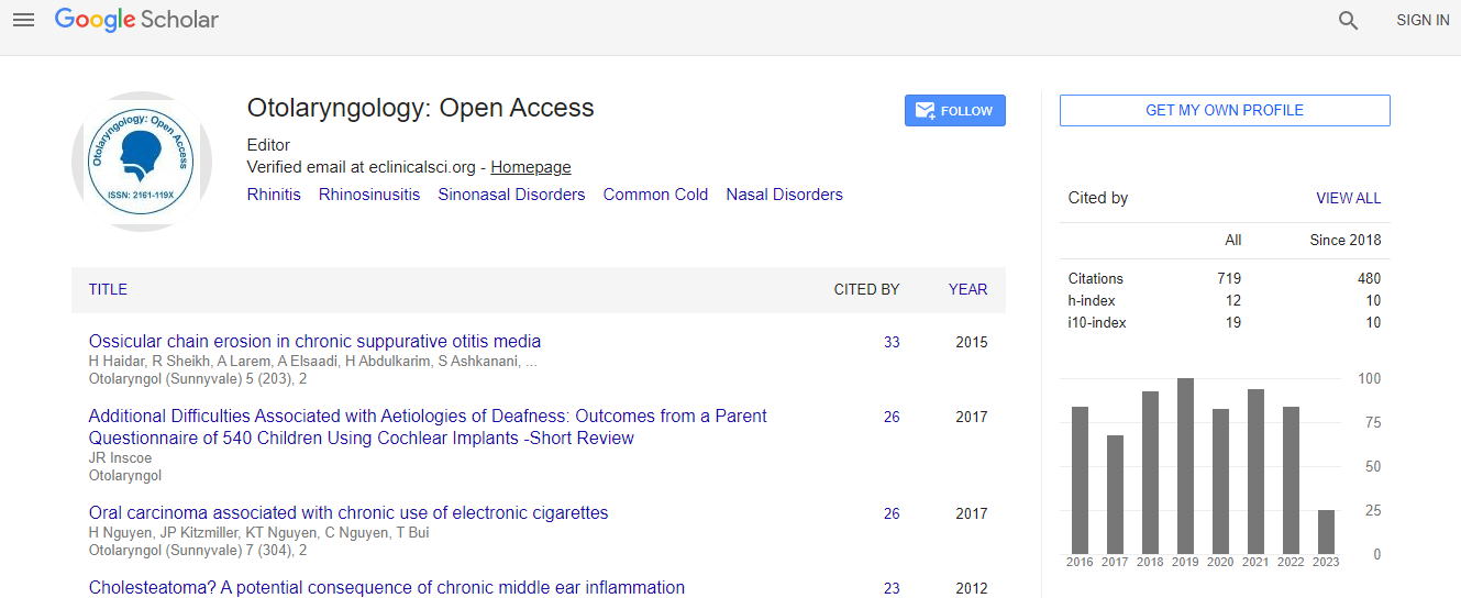
Google Scholar citation report
Citations : 925
Otolaryngology: Open Access received 925 citations as per Google Scholar report
Otolaryngology: Open Access peer review process verified at publons

Indexed In
- Index Copernicus
- Google Scholar
- Sherpa Romeo
- Open J Gate
- Genamics JournalSeek
- RefSeek
- Hamdard University
- EBSCO A-Z
- OCLC- WorldCat
- Publons
- Geneva Foundation for Medical Education and Research
- ICMJE
Useful Links
Recommended Journals
Related Subjects
Share This Page
Automatic digital image capture and archiving: Where and why we drop the ball, and how we can pick it up again
International Conference and Exhibition on Otolaryngology
David D Pothier
ScientificTracks Abstracts: Otolaryngology
Abstract
Otolaryngology involves the use of a number of sophisticated instruments to examine the upper aerodigestive system and ears. Although it is easily possible to take digital images with most of the devices used to examine the ear, nose and throat, this is seldom undertaken in current clinical practice, with drawings/diagrams usually being used to record clinical appearances. Drawings have, however, been shown conclusively to be almost completely useless in the recording of pathology of the tympanic membrane and larynx. With the development of cheap digital auriscopes, clinical images of the tympanic membrane can be captured very easily. Unfortunately archiving remains very problematic. Traditionally, taking images to a storage medium meant images had to be manually labeled and stored with patient metadata entered for each image. This made real-time image capture impractical for clinical purposes. An Automatic Image Capture and Archiving System (AICAS) allows a clinician to capture images from any USB input i.e. digital auriscope, output from endoscopic stack etc. and then automatically stores the images in a central database under the details of the patient being examined. On review of the patient, all previous images are retrieved for comparison and new images can be added to the record automatically. An AICAS system allows for the accurate recording of pathology using digital images which, in addition to providing an accurate record, allows for transparent audit, patient involvement in treatment decisions, additional educational opportunities and the opportunity to undertake large scale cross sectional and longitudinal studies of pathologies at no cost, using archived images. In this lecture, an AICAS system is described and its introduction into clinical practice is detailed. All Otolaryngologists became familiar with the system within 90 minutes of its introduction and, after one week, the use of the system added a median of 11 seconds to each consultation. Feedback was positive across all levels of use and the system remains in place in both hospitals.Biography
Dr Pothier attended medical school at the University of Cape Town, South Africa. He then undertook his Otolaryngology training in London and the South West of England. Fellowship trained in Neurotology in Toronto, Canada where he now works as anOtologist/Neurotologist with an interest in middle ear and vestibular disorders, as well as Health Informatics. He has a primary research interest in bilateral vestibular loss. He has published over 80 peer-reviewed articles and has a Bacon number of 4.