Our Group organises 3000+ Global Conferenceseries Events every year across USA, Europe & Asia with support from 1000 more scientific Societies and Publishes 700+ Open Access Journals which contains over 50000 eminent personalities, reputed scientists as editorial board members.
Open Access Journals gaining more Readers and Citations
700 Journals and 15,000,000 Readers Each Journal is getting 25,000+ Readers
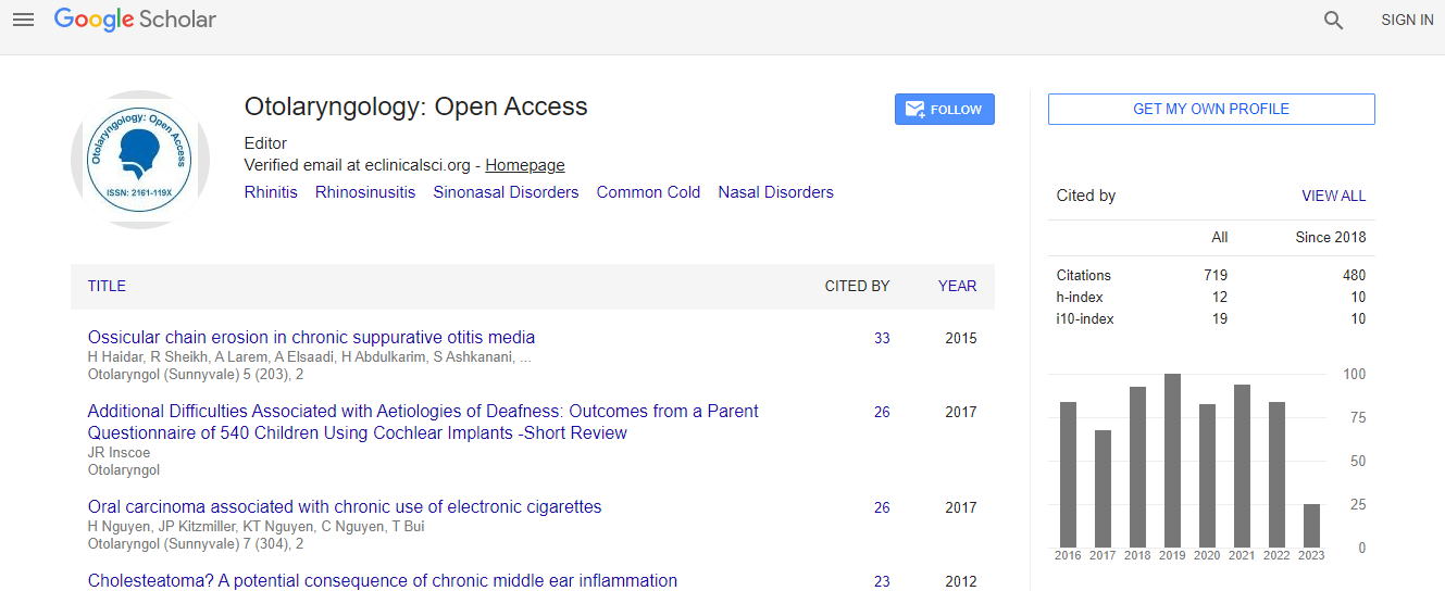
Google Scholar citation report
Citations : 925
Otolaryngology: Open Access received 925 citations as per Google Scholar report
Otolaryngology: Open Access peer review process verified at publons
Indexed In
- Index Copernicus
- Google Scholar
- Sherpa Romeo
- Open J Gate
- Genamics JournalSeek
- RefSeek
- Hamdard University
- EBSCO A-Z
- OCLC- WorldCat
- Publons
- Geneva Foundation for Medical Education and Research
- ICMJE
Useful Links
Recommended Journals
Related Subjects
Share This Page
Application of advanced virtual reality and 3D-computer assisted technologies in NESS
International Conference on Aesthetic Medicine and ENT
Ivica Klapan
The School of Medicine University of Zagreb, Croatia The Schools of Medicine J J Strossmayer University in Osijek, Croatia Klapan Medical Group Polyclinic, Croatia
Posters & Accepted Abstracts: Otolaryngology
Abstract
In the modern-day world medical technology, NESS-systems represent the technique with highly precise, extremely small navigation instruments which guides the surgeon through the software, provides the most flexible or-setup, with automatic recognition of the surgeon├ó┬?┬?s intent during the procedure, and with no need to press a button, but with some functional limitations. If surgeons would need additional information (e.g., how deep, and where the pathologic process invaded standard normal mucosal layer inside the sinus├ó┬?┬Ł etc.), do they have appropriate, and sufficient support given by NESS, just even in very simple cases? The answer is ├ó┬?┬?no├ó┬?┬Ł! But with additional application of several (semi) automatic tools (e.g. wave-propagation, skeletonbased approaches, and methods based on depth-maps), developed as simulated spaces (artificial reality), it is possible to provide appropriate support in OR (detection of regions of interest, structural and functional analyses, data-driven visualization techniques for data exploration). From the very beginning of my 3D-CA-NESS/1994, and tele3D-CA-NESS/1998, 3D-image analysis and processing, tissue modeling, and virtual endoscopy/surgery, represented a basis for various realistic simulations in standard-FESS. The possibility of exact preoperative, non-invasive visualization of the spatial relationships of anatomic and pathologic structures, including extremely fragile ones, size and extent of pathologic process, and of precisely predicting the course of surgical procedure, allowed me considerable advantage in the preoperative examination of the patient and to reduce the risk of intraoperative complications (all this by use of different VR-methods). Real-time-VR-technology will update the 3D-graphical visualization of the patient├ó┬?┬?s anatomy, providing a highly useful and informative visualization of the regions of interest, thus bringing advancement in defining the geometric information on anatomical contours of 3D-humanhead models by the transfer of so-called image pixels to contour pixels.Biography
Email: telmed@mef.hr

 Spanish
  Chinese
  Russian
  German
  French
  Japanese
  Portuguese
  Hindi
Hindi 
