Our Group organises 3000+ Global Conferenceseries Events every year across USA, Europe & Asia with support from 1000 more scientific Societies and Publishes 700+ Open Access Journals which contains over 50000 eminent personalities, reputed scientists as editorial board members.
Open Access Journals gaining more Readers and Citations
700 Journals and 15,000,000 Readers Each Journal is getting 25,000+ Readers
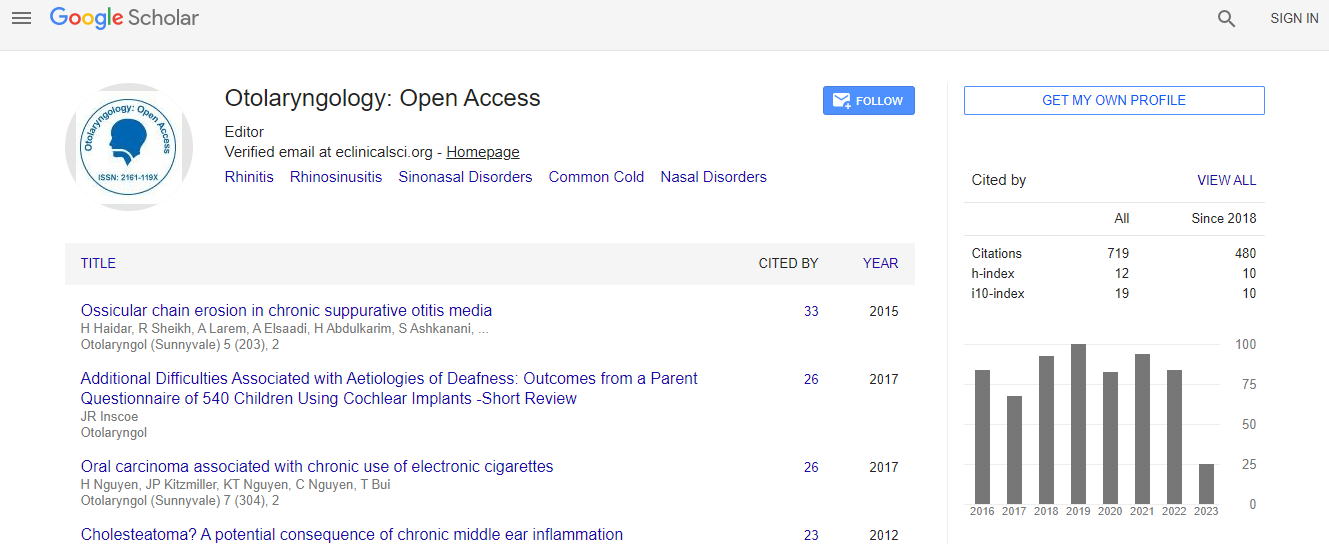
Google Scholar citation report
Citations : 925
Otolaryngology: Open Access received 925 citations as per Google Scholar report
Otolaryngology: Open Access peer review process verified at publons
Indexed In
- Index Copernicus
- Google Scholar
- Sherpa Romeo
- Open J Gate
- Genamics JournalSeek
- RefSeek
- Hamdard University
- EBSCO A-Z
- OCLC- WorldCat
- Publons
- Geneva Foundation for Medical Education and Research
- ICMJE
Useful Links
Recommended Journals
Related Subjects
Share This Page
An imaging technology for intra-operative surgical margin assessment in oral and head and neck cancers (OSCC)
3rd International Conference and Exhibition on Rhinology & Otology
Maie St John
David Geffen School of Medicine at UCLA, USA
Keynote: Otolaryngology
Abstract
Oral and Head and neck squamous cell carcinoma (OSCC) is the sixth most common cancer in the world. The primary management of OSCC relies on complete surgical resection of the tumor. However, the establishment of margin-free resection is often difficult given the devastating side effects of aggressive surgery and the anatomic proximity to vital structures such as the carotid artery and the spinal cord. Positive margin status is associated with significantly decreased survival. Currently, it is the surgeon�s fingers that determine where the tumor cuts are made, by palpating the edges of the tumor. Accuracy varies widely based on the experience of the surgeon and the location and type of tumor. Efficacy is further confounded by the risk of damage to adjacent vital structures, which limit resection margins. The goal of this proposal is to evaluate a novel, non-invasive, imaging system based on Dynamic Optical Contrast Imaging (DOCI) that has been developed to differentiate between cancerous and normal tissue intra-operatively using OSCC as the model. The imaging system is based on a novel realization of temporally dependent measurements of tissue auto-fluorescence that allow the acquisition of specific tissue properties over a large field of view. This system is optimized such that it can be used by surgeons at the time of cancer resection surgery to gather quantitative information on margins of malignancies and has been extensively validated in ex vivo OSCC samples. Companion histology has verified the sensitivity and specificity of the technique. This intra-operative instrument would be the first of its kind, giving us the potential to significantly improve the sensitivity and accuracy of determining true OSCC margins thus enabling the surgeon to save healthy tissue and improve patient outcomes.Biography
Maie St John is an academic head & neck surgeon with a passion for education. She holds the Pearlman endowed Chair in Otolaryngology/Head and Neck Surgery and is the Co-Director of the UCLA Head and Neck Cancer Program. The focus of her clinical work is the treatment of head and neck tumors. As chair of the Curriculum Committee in the Department of Head and Neck Surgery at UCLA, she developed a critical and comprehensive education program that prepares our graduates for board certification. Recently, she also received the UCLA Health System Humanism Award, as one of the top 10 most humanistic physicians at UCLA. She is an active member of several professional societies, including: the American Academy of Otolaryngology-Head and Neck Surgery, the American Head and Neck Society, the Los Angeles Biomedical Research Institute, the Triological Society and the American Association for Cancer Research.
Email: mstjohn@mednet.ucla.edu

 Spanish
  Chinese
  Russian
  German
  French
  Japanese
  Portuguese
  Hindi
Hindi 
