Our Group organises 3000+ Global Conferenceseries Events every year across USA, Europe & Asia with support from 1000 more scientific Societies and Publishes 700+ Open Access Journals which contains over 50000 eminent personalities, reputed scientists as editorial board members.
Open Access Journals gaining more Readers and Citations
700 Journals and 15,000,000 Readers Each Journal is getting 25,000+ Readers
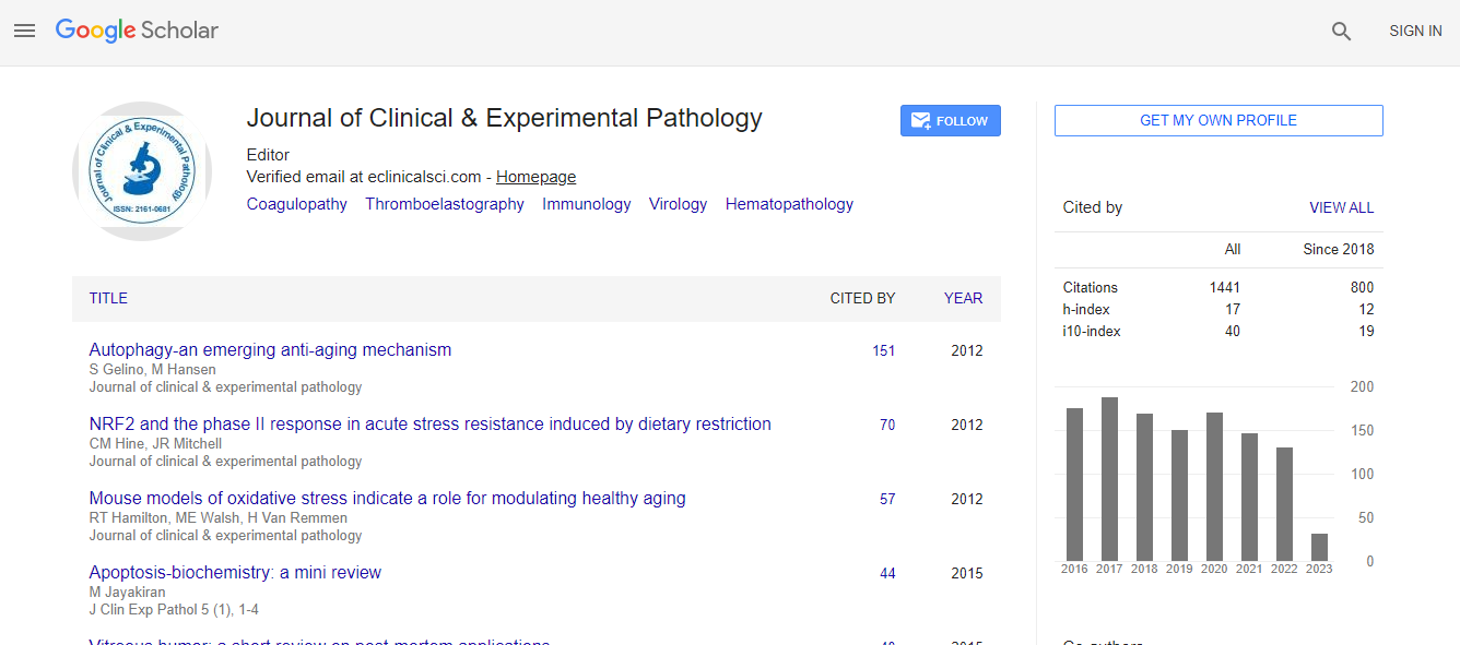
Google Scholar citation report
Citations : 2975
Journal of Clinical & Experimental Pathology received 2975 citations as per Google Scholar report
Journal of Clinical & Experimental Pathology peer review process verified at publons
Indexed In
- Index Copernicus
- Google Scholar
- Sherpa Romeo
- Open J Gate
- Genamics JournalSeek
- JournalTOCs
- Cosmos IF
- Ulrich's Periodicals Directory
- RefSeek
- Directory of Research Journal Indexing (DRJI)
- Hamdard University
- EBSCO A-Z
- OCLC- WorldCat
- Publons
- Geneva Foundation for Medical Education and Research
- Euro Pub
- ICMJE
- world cat
- journal seek genamics
- j-gate
- esji (eurasian scientific journal index)
Useful Links
Recommended Journals
Related Subjects
Share This Page
A validation study of WSI-based primary diagnosis for malignant lymphoma
2nd International Conference on Digital Pathology & Image Analysis
Tomoo Itoh
Kobe University Hospital, Japan
Keynote: J Clin Exp Pathol
Abstract
Background: The digital pathology is an emerging technology, and its usage on routine practices is spreading worldwide rapidly. Very recently, FDA allowed marketing of first whole slide imaging (WSI) system for digital pathology, which enables us use the system even for primary diagnosis. This epoch-making achievement owes a lot to scientific evidences indicated that WSI is eligible for making accurate pathological diagnoses. However, those studies typically targeting small specimens alone and the cases requiring immunohistochemistry or special staining, such as malignant lymphoma, were excluded in many studies. Objective: To provide an evidence of usability of WSI diagnosis for primary diagnosis of malignant lymphoma compared to conventional glass slide diagnosis and optical microscope. Design: The cases of malignant lymphoma were retrieved from our case collection. The all slide glasses, including H&E and immunohistochemistry were digitized using a WSI scanner, NanoZoomer RS (Hamamatsu), with X40 magnification, and a well-trained pathologist for lymphoma diagnosis had reviewed and made diagnosis for the digitized cases with more than 2 months of washout time interval. Discrepancies between microscope slide and WSI diagnosis were classified into three categories; concordance, major discrepancy (defined as ones associated with significant difference in clinical treatment), and minor discrepancy (defined as ones associated with no significant difference in clinical treatment). Result: At the time of writing this abstract, the study was still ongoing. Tentative data showed excellent concordance rate, over than 95%, and which was much better than we expected. Conclusion: WSI is applicable for primary diagnosis of malignant lymphoma, if we make diagnoses with combination of adequate clinical information, H&E morphology, and immunohistochemistries.Biography
Tomoo Itoh has completed his PhD at Hokkaido University Graduate School of Medicine and presently he is a Professor and Deputy Director of Diagnostic Pathology at Kobe University Hospital, Japan. He is a Board Certified Member of the Japanese Society of Pathology and Board Certified Member of the Japanese Society of Clinical Cytology. He was President of 15th Annual Meeting of Japanese Society of Digital Pathology held in Kobe in 2016, and now one of the core members of the Society.

 Spanish
  Chinese
  Russian
  German
  French
  Japanese
  Portuguese
  Hindi
Hindi 
