Our Group organises 3000+ Global Conferenceseries Events every year across USA, Europe & Asia with support from 1000 more scientific Societies and Publishes 700+ Open Access Journals which contains over 50000 eminent personalities, reputed scientists as editorial board members.
Open Access Journals gaining more Readers and Citations
700 Journals and 15,000,000 Readers Each Journal is getting 25,000+ Readers
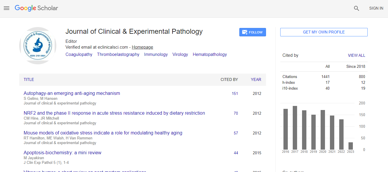
Google Scholar citation report
Citations : 2975
Journal of Clinical & Experimental Pathology received 2975 citations as per Google Scholar report
Journal of Clinical & Experimental Pathology peer review process verified at publons
Indexed In
- Index Copernicus
- Google Scholar
- Sherpa Romeo
- Open J Gate
- Genamics JournalSeek
- JournalTOCs
- Cosmos IF
- Ulrich's Periodicals Directory
- RefSeek
- Directory of Research Journal Indexing (DRJI)
- Hamdard University
- EBSCO A-Z
- OCLC- WorldCat
- Publons
- Geneva Foundation for Medical Education and Research
- Euro Pub
- ICMJE
- world cat
- journal seek genamics
- j-gate
- esji (eurasian scientific journal index)
Useful Links
Recommended Journals
Related Subjects
Share This Page
A multilocular simple bone cyst associated with cemento-osseous dysplasia
5th International Conference on Pathology
Sung Yong Han, Morhaf Sadek, Ibrahim Zakhary and Brent Accurso
University of Detroit Mercy School of Dentistry, USA Oral Pathology Consultants PLLC, USA
Posters & Accepted Abstracts: J Clin Exp Pathol
Abstract
A 49 year old African American female was referred to the Oral and Maxillofacial Surgery Clinic at the University of Detroit Mercy, School of Dentistry for evaluation of a large, right mandibular radiolucency. Clinical examination revealed an asymptomatic slight buccal expansion of the right mandible. All teeth tested vital. Panoramic radiography revealed a large well defined, multilocular radiolucent lesion spanning from the apex of tooth #27 to the ascending ramus distal to tooth #32. Other radiopaque lesions with radiolucent margins were noted at the apices of the mandibular incisors, left canine, first premolar and first molar. Computed tomography revealed a non specific expansile cystic lesion within the right mandibular body surrounding the roots of the right mandibular first through third molars as well as partially surrounding the roots of the right mandibular canine and the first and second premolars. Exploration of the lesion was performed under local anesthesia. Aspiration of the lesion yielded a yellowish fluid and the cavity that was surgically explored appeared hollow with a thin soft tissue lining. The H&E stain histopathology showed a mixture of loose fibrovascular connective tissue, fragments of vital bone, a mild infiltrate of chronic inflammatory cells and extravasated erythrocytes. The histopathology, in conjunction with the surgical findings and radiography were consistent with a diagnosis of a simple bone cyst with concomitant cemento-osseous dysplasia. Observation of the lesion will be continued until complete healing is radiographically confirmed and outcome of the lesion will be reported in the future.Biography
Sung Yong Han is currently a 4th year Dental Student at the University of Detroit, Mercy School of Dentistry, Detroit, MI.
Email: jminden08@gmail.com

 Spanish
  Chinese
  Russian
  German
  French
  Japanese
  Portuguese
  Hindi
Hindi 
