Research Article Open Access
Variation in to Neural Physiology and Anatomy Due to Diet Related Factors as a Possible Cause to Neurodegeneration
Kamala Govindaraju1, Jaya Gopalkrishna1, Jennifer Thankachan Thomas1, Nisha Narayan Moger1 and Maulishree Agrahari1,2*1PES Institute of Technology, Bangalore, India
2University of Calgary, Canada
- Corresponding Author:
- Maulishree Agrahari
University of Calgary, Canada
E-mail: kamala.037@gmail.com
Received date: May 08, 2012; Accepted date: June 25, 2012; Published date: June 27, 2012
Citation: Govindaraju K, Gopalkrishna J, Thomas JT, Moger NN, Agrahari M (2012) Variation in to Neural Physiology and Anatomy Due to Diet Related Factors as a Possible Cause to Neurodegeneration. J Biotechnol Biomater 2:141. doi:10.4172/2155-952X.1000141
Copyright: © 2012 Govindaraju K, et al. This is an open-access article distributed under the terms of the Creative Commons Attribution License, which permits unrestricted use, distribution, and reproduction in any medium, provided the original author and source are credited.
Visit for more related articles at Journal of Biotechnology & Biomaterials
Abstract
Neurodegenerative disorders have been linked to an array of external factors ranging from atmospheric pollutants to diet related additives. In our study we have investigated BMAA a neurotoxin produced by cyanobacteria, which gets an easy entry into human food chain via sea food and water, Aluminium which is believed to be present more in bottled water in comparison to tap water; and MSG, a derivative of glutamate; extensively used as flavour enhancer. LD50 for the three chemicals used in this study were determined. Our results suggest that BMAA leads to a significant loss of long term memory, reduction in geotaxic response, and reduction in both, fertility and life span. Short term memory was not significantly affected by BMAA. Aluminium deposits caused loss of long and short term memory and reduction in geotaxic response. MSG caused reduction in short term memory. In combination with BMAA, MSG produced reduction in all the parameters. Dementia has a direct bearing on quality of life. Dementia can set in as early as 40 years of age in humans. This has been attributed to life-style and stress related factors. The present study establishes a direct cause and effect relationship between dementia, stress and diet. These effects are established when Drosophila melanogaster are treated with neurotoxins, singly or in combinations. Experiments are in progress to understand the mechanisms that underlie these processes.
Keywords
Drosophila melanogaster; Neurodegeneration; Alzheimer’s and Parkinson’s diseases
Introduction
Alzheimer’s and Parkinson’s diseases and Amyotrophic Lateral Sclerosis (ALS) are the most common age-related neurodegenerative diseases worldwide. There are several environmental factors which are responsible for these neurological disorders [1]. Our studies mainly concentrates on 3 different chemicals namely i. BMAA ii. aluminium iii. MSG and also we have studied their combination. In the 1950s a strange neurological problems was reported on the island of Guam in the Western Pacific. Histological observations of their brain showed damages similar to what is seen in Alzheimer’s disease, though the disease was distinct from Alzheimer’s. This was termed as ALS-PDC (Amyotrophic Lateral Sclerosis Parkinsonism-Dementia Complex). At first, ALS-PDC was believed to be genetic in nature. Previous cytotoxic studies flourished the awareness of high ALS incidence on the South Pacific island of Guam and in conjunction with dietary epidemiological observations, these studies have allowed for the isolation of a potential neurotoxin, β-N-methylamino-l-alanine (BMAA) [2,3]. BMAA is a neurotoxic, non-lipophilic, non-protein amino acid produced by cyanobacteria. In the Guam ecosystem BMAA is produced by cyanobacteria of the genus Nostoc, which are root symbionts of cycads, and is then biomagnified in cycad seeds and in Marianas flying foxes (Pteropus mariannus) that forage on them [4]. Consumption of flying foxes by the Chamorro people has been implicated as a delivery mechanism for relatively high doses of BMAA in the Chamorro diet [2]. BMAA occurs not only as a free amino acid in the ecosystem but also can be released from a protein-bound form by acid hydrolysis [5]. BMAA has been detected in both free and protein-bound form in the brain tissues of Chamorro patients who died from ALS/PDC [5]. Beta- N-methylamino-L-alanine (L-BMAA) is a neurotoxic non-protein amino acid that is produced by cyanobacteria, a blue-green algae that is common to many lakes, oceans, and soils, and is found in Cycascircinalis seeds. It has previously been associated with the neurological disease amyotrophic lateral sclerosis-Parkinson dementia complex (ALS-PDC) in an indigenous population in Guam [2,4]. Consumption of tortillas made from cycad-seed flour and of flying foxes (Mariana fruit bats, a “traditional delicacy” in Guam) that feed on cycad fruit has been implicated as the source of L-BMAA exposure. Proteinbound and free L-BMAA has been shown to bio concentrate in the Guam ecosystem (bacteria, plants, animals, and human tissues) and detectable levels have been measured in brain tissue of people in Guam who suffered from dementia-related disorders. Because it is present in blue-green algae from a variety of water sources around the world and samples of Baltic Sea and oceanic blooms, significant quantities may be released into the world’s oceans and water supplies [6-9]. In case of Al some toxicity can be traced to deposition in bone and the central nervous system as competes with calcium for absorption and may contribute to the reduced skeletal mineralization (osteopenia). In very high doses, aluminium can cause neurotoxicity, and is associated with altered function of the blood-brain barrier [10,11]. Aluminium enters the environment through deodorants, purified water, medicines, utensils, foils, tooth paste and capsule covers. Monosodium glutamate, also known as sodium glutamate and MSG, is a sodium salt of glutamic acid, a naturally occurring non-essential amino acid. It is used as a food additive and is commonly marketed as a flavour enhancer. Some studies have shown its link to neurodegenerative disorders. Since these neurotoxins enters the environment through various ways and causes the neurological disease. Our studies mainly help to show the effects of these neurotoxins. The main effects which have been studied are i. memory ii. Response and iii. Fertility. We have used drosophila melanogester as our model organism [12,13]. And the experiment is conducted on the basis of age (newly hatched files). LD50 studies have been conducted for aluminium and MSG.
Materials and Methods
Drosophila breeding and culturing
Canton S (CS) flies were raised on grape –agar media at room temperature (37 °C) under ambient light. For BMAA and other amino acid feeding experiments, BMAA(Sigma-Aldrich, St. Louis, U.S.), aluminium (aluminium sulphate), MSG (normal ajjinomoto ) were dissolved in double distilled water and mixed into the yeast paste. The different concentration of BMAA, aluminium and MSG was mixed with dry yeast and distilled water. CS flies newly hatched-1day flies were separated around ten files were used for each concentration.
LD50
LCt50 (Lethal Concentration & Time) of a toxin, radiation, or pathogen is the dose required to kill half the members of a tested population after a specified test duration. LD50 figures are frequently used as a general indicator of a substance’s acute toxicity. LD50 studies were conducted foe Aluminium and MSG. The flies (newly hatchedone old) were fed with different concentration of aluminium and MSG along with yeast paste. And the data was noted.
Olfactory test
The olfactory test measures the learning and memory abilities of the flies. Both long term and short term was tested. Short term memory: The newly hatched flies were fed with neurotoxin for 48 hours. The flies were trained using the T-shaped tubes. The blue tube was associated with food (yeast paste) and was keeping in the light for 5min and then transferred to the yellow tube which was not associated with food. Same was repeated for three times. And the flies were rested for 30min and then the memory of the flies was tested by allowing each fly at a time. T-shaped testing tube was made by covering one side with blue colour and the other side with yellow colour. If the fly move toward the blue then it is a good learner, if it moves towards yellow then it is a bad learner. Long term memory test: the normal flies were trained and tested after 24 hours and the good learners and bad learners were separated and the good learners were fed with a neurotoxin for 48 hours and then tested.
Phototaxis aversion assay
The phototaxic suppression assay measures the learning and memory ability of flies. It was carried out a with a modified T-shape tube one side of the tube was covered with the dark and other side was modified in such a way that single beam of light was passed through and Filter paper soaked with 1mM quinine hydrochloride solution was placed in the lighted (narrow beam light) [13,14]. The whole set was carried out in a square box. The short term and long term memory was test. The flies were fed with the neurotoxin and tested after 48 hours. The flies were allowed to pass through the T-shape tube for 5mins. The flies were in rest for 5mins. The same was repeated three times. And the flies were rested for 30min and then the memory of the flies was tested by allowing each fly at a time. T-shaped testing tube was made by covering one side with dark and the other side with light. If the fly move toward the dark then it is a good learner, if it moves towards light then it is a bad learner. Long term memory test: the normal flies were trained and tested after 24 hours and the good learners and bad learners were separated and the good learners were fed with a neurotoxin for 48 hours and then tested.
Geotaxic response
The geotaxis assay (climbing assay) was performed as described response of the files. Flies empty borsil glass bottle of height 35 cm. In the behaviour assay, flies were gently tapped to the bottom of the vial and then allowed to crawl. In response to tapping (gentle agitation), flies crawl/climb upward against gravity [12,15]. The test was conducted for the normal flies and the height was taken at 1min the test was conducted for 10 flies and the average time and height was noted. The normal files were fed with different concentration of neurotoxins for 48 hours. Then each fly were tested passing the files to the vertical bottle and the time was given for 1min .Any flies that died during the course of the assay were eliminated and the data points associated with dead flies were disregarded.
Fertility test for normal and BMAA fed female and male flies
Fertility test was conducted to determine the number of eggs,larvae, cocoons and number of flies hatched. The test was conducted for normal and BMAA fed flies. Two females and three males were put in the culture bottle set up. Each day the flies are transferred to another new bottle and same is repeated for 4 days. Each day number of eggs, larvae, cocoons and flies are counted. And the results of normal are compared with the neurotoxin fed flies. And also the different stages and number of days of life cycle was calculated.
Results
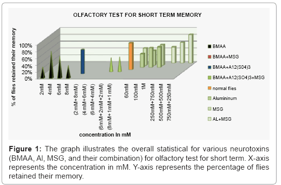
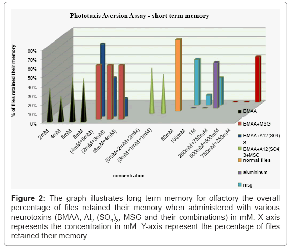
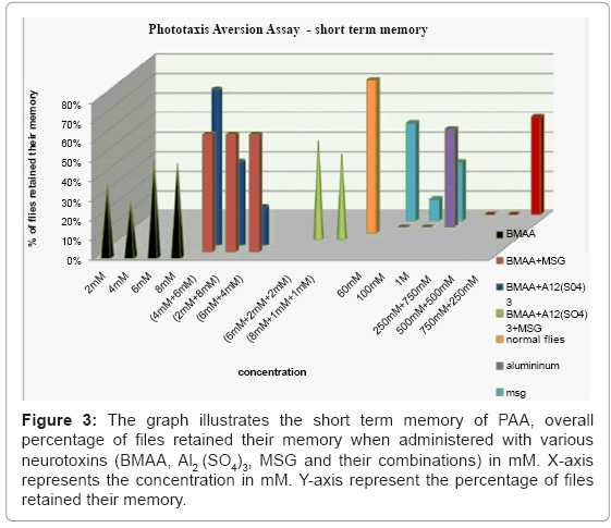
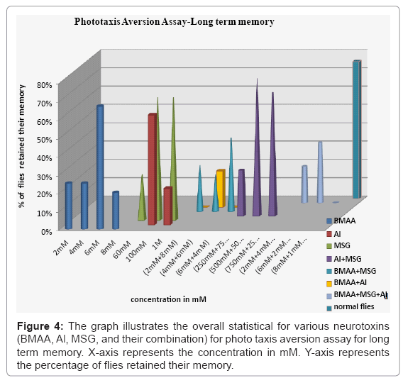
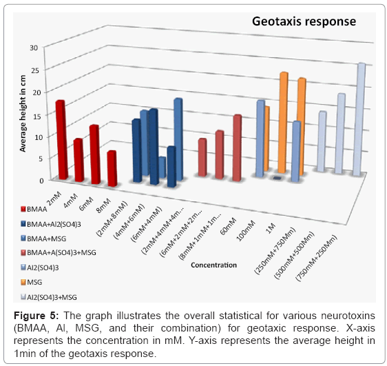
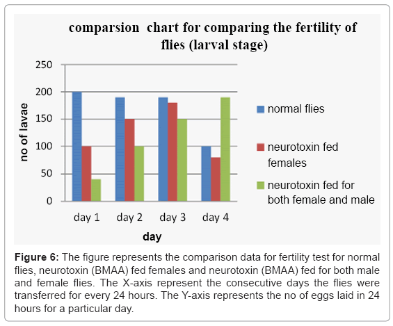
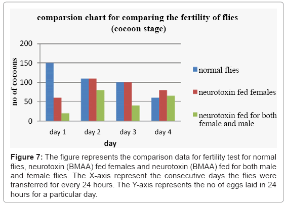
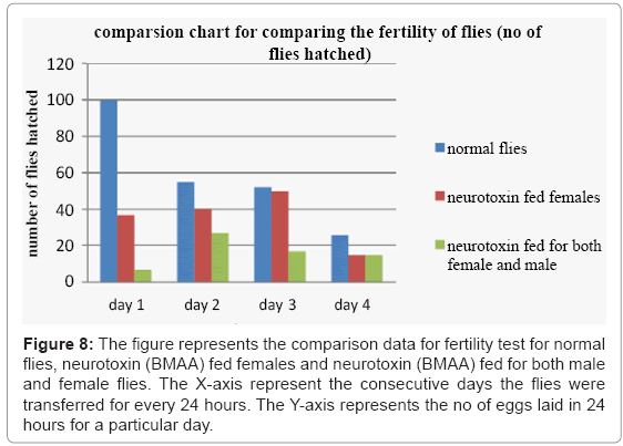
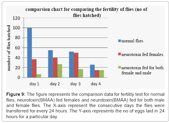
The fruit fly Drosophila has been used to study human neurodegenerative diseases and these studies have contributed greatly to our understanding of the mechanisms of neurodegeneration and environmental factors which are responsible for them. Specifically, Drosophila has been shown to be an ideal model to quantitatively measure motor coordination and learning and memory, as flies have distinctive behaviours that are stereotypic and quantifiable, such as geotaxic and phototaxic behaviour. Geotaxis refers to the climbing behaviour of flies and their natural tendency to climb against gravity, where a reduced climbing behaviour indicates reduced motor coordination. Phototaxis refers to the instinctive behaviour of flies to move toward light. The olfactory system is the sensory system used for olfaction, or the sense of smell, the olfactory test investigates the behaviour of the flies to move toward the food. LCt50 (Lethal Concentration & Time) of a toxin, radiation, or pathogen is the dose required to kill half the members of a tested population after a specified test duration we performed the LD50 studies for MSG and Aluminium (Table 1-4). In order to test the specific effects of the three neurotoxins (BMAA, Aluminium and MSG) on central nervous system function, we examined the learning and memory abilities of flies upon neurotoxin treatment using the phototaxic aversion assay and olfactory test .Flies are instinctively phototaxic, but quinine, on the other hand, induces avoidance behaviour and is frequently used as a negative reinforcer. In the aversive phototaxic suppression assay, flies were individually placed into a T- tube and allowed to choose between a lighted and a darkened side. Filter paper is soaked with quinine solution of concentration 1mM and placed into the lighter side of the T-Tube such that the quinine provided an aversive association. During the conditioning phase, flies learn to suppress their instinctively phototaxic behaviour in order to avoid quinine. Hence the flies were first fed with neurotoxin (48 hours). Then flies were trained and tested for their memory. There was maximum effects on (BMAA + Aluminium sulphate) of (2mM + 8mM) and (6mM + 4mM) and (BMAA + Aluminium sulphate + MSG) of (8mM + 1mM + 1mM) fed flies for long term memory (Figure 4). In short term memory the maximum effect was seen in aluminium (60mM,100mM), BMAA(4mM), (al + MSG) ((250 + 750), (500Mm + 500mM)) and MSG (100mM) fed flies (Figure 3). In olfactory test, flies were individually placed into a T- tube and allowed to choose between a blue and a yellow side. Blue tube was associated with food and yellow tube was not associated with food. During the conditioning phase, flies learn to move towards the blue tube due to smell although they have the affinity to move towards the light, in this conditioning phase the flies are trained. In case of long term memory All tests conducted showed loss of long term memory, all combinations of MSG + BMAA, 4mM BMAA, (4mM + 6mM) and (6mM + 4mM) and (4mM + 6mM) of BMAA +Al2 (SO4)3 and (8mM + 1mM + 1mM) and (2mM + 4mM + 4mM) BMA + MSG + Al2 (SO4)3 resulted in memory loss of all flies involved in the test (Figure 2). In short term memory was not that affected. The 2mM BMAA showed the memory loss and also the combinations showed the substantial memory loss (Figure 1). We examine the locomotors functioning using the geotaxic response. The flies were fed with different concentration of neurotoxins. The results are Aluminum sulphate, BMAA, MSG and there combinations cause locomotor dysfunction which leads to reduction in geotaxic response (Figure 5). The t-test for two difference means was performed and thus it was proved that these neurotoxins cause the locomotor dysfunction. We examine the fertility of the flies by feeding the female flies with the neurotoxin for 24 hours and then we made them to mate with normal male and fed them with normal yeast paste and also we made female flies mate with male flies by feeding them with 4mM concentration of neurotoxin BMAA. Then we compare the results with the normal flies. We observed that the neurotoxic flies deviated from the normal behaviour and also showed reduced production in eggs (Figure 6), larvae (Figure 7), cocoons (Figure 8) and flies hatched (Figure 9).
| Concentration (mM) |
No. of flies | DAY 1 | DAY 2 | DAY 3 | DAY 4 |
|---|---|---|---|---|---|
| 0.5 | 10 | 10 | 9 | 9 | 8 |
| 1.0 | 9 | 9 | 9 | 9 | 9 |
| 1.5 | 7 | 7 | 7 | 7 | 7 |
| 2.0 | 12 | 12 | 10 | 7 | 4 |
| 3.0 | 7 | 7 | 7 | 7 | 7 |
| 3.5 | 7 | 7 | 7 | 7 | 7 |
| 4.0 | 9 | 9 | 9 | 9 | 9 |
| 4.5 | 12 | 12 | 12 | 12 | 12 |
| 5.0 | 9 | 9 | 9 | 9 | 9 |
| 7.0 | 9 | 9 | 9 | 9 | 9 |
| 10.0 | 10 | 10 | 10 | 10 | 10 |
Table 1: LD50 data for Al2 (SO4)3 in mM.
| Concentration (mM) |
No. of flies | DAY 1 | DAY 2 | DAY 3 | DAY 4 |
|---|---|---|---|---|---|
| 20 | 7 | 7 | 7 | 7 | 7 |
| 30 | 8 | 8 | 8 | 8 | 8 |
| 40 | 7 | 7 | 7 | 6 | 6 |
| 50 | 6 | 6 | 5 | 5 | 5 |
| 60 | 8 | 8 | 8 | 8 | 8 |
| 70 | 8 | 8 | 7 | 7 | 7 |
| 80 | 7 | 7 | 6 | 6 | 6 |
| 90 | 9 | 9 | 9 | 8 | 8 |
| 100 | 9 | 9 | 8 | 8 | 8 |
| 110 | 7 | 7 | 7 | 7 | 7 |
Table 2: LD50 data for Al2 (SO4)3 in mM.
| Concentration (mM) |
No. of flies | DAY 1 | DAY 2 | DAY 3 | DAY 4 |
|---|---|---|---|---|---|
| 0.5 | 10 | 10 | 9 | 8 | 7 |
| 1.0 | 10 | 10 | 10 | 10 | 10 |
| 1.5 | 7 | 7 | 7 | 7 | 7 |
| 2.0 | 12 | 12 | 12 | 9 | 9 |
| 2.5 | 20 | 20 | 19 | 18 | 17 |
| 3.0 | 9 | 9 | 9 | 9 | 9 |
| 3.5 | 11 | 11 | 11 | 11 | 11 |
| 4.0 | 14 | 14 | 13 | 12 | 12 |
| 4.5 | 8 | 8 | 8 | 8 | 8 |
| 5.0 | 10 | 10 | 10 | 10 | 10 |
| 7.0 | 12 | 12 | 12 | 8 | 7 |
| 10.0 | 14 | 14 | 13 | 13 | 13 |
Table 3: LD50 data for MSG in mM.
| Concentration (mM) |
No. of flies | DAY 1 | DAY 2 | DAY 3 | DAY 4 | DAY 5 |
|---|---|---|---|---|---|---|
| 20 | 6 | 6 | 6 | 6 | 6 | 5 |
| 40 | 7 | 7 | 7 | 7 | 7 | 7 |
| 60 | 6 | 6 | 6 | 6 | 6 | 6 |
| 80 | 9 | 9 | 9 | 9 | 9 | 9 |
| 100 | 6 | 6 | 6 | 6 | 6 | 6 |
| 120 | 6 | 6 | 6 | 6 | 6 | 6 |
| 140 | 9 | 9 | 9 | 9 | 9 | 9 |
| 160 | 12 | 12 | 11 | 11 | 11 | 11 |
| 180 | 6 | 6 | 6 | 6 | 6 | 6 |
| 200 | 9 | 9 | 9 | 9 | 9 | 9 |
Table 4: LD50 data for MSG in mM.
Figure 2: The graph illustrates long term memory for olfactory the overall percentage of files retained their memory when administered with various neurotoxins (BMAA, Al2 (SO4)3, MSG and their combinations) in mM. X-axis represents the concentration in mM. Y-axis represent the percentage of files retained their memory.
Figure 3: The graph illustrates the short term memory of PAA, overall percentage of files retained their memory when administered with various neurotoxins (BMAA, Al2 (SO4)3, MSG and their combinations) in mM. X axis represents the concentration in mM. Y-axis represent the percentage of files retained their memory.
Figure 6: The figure represents the comparison data for fertility test for normal flies, neurotoxin (BMAA) fed females and neurotoxin (BMAA) fed for both male and female flies. The X-axis represent the consecutive days the flies were transferred for every 24 hours. The Y-axis represents the no of eggs laid in 24 hours for a particular day.
Figure 7: The figure represents the comparison data for fertility test for normal flies, neurotoxin (BMAA) fed females and neurotoxin (BMAA) fed for both male and female flies. The X-axis represent the consecutive days the flies were transferred for every 24 hours. The Y-axis represents the no of eggs laid in 24 hours for a particular day.
Figure 8: The figure represents the comparison data for fertility test for normal flies, neurotoxin (BMAA) fed females and neurotoxin (BMAA) fed for both male and female flies. The X-axis represent the consecutive days the flies were transferred for every 24 hours. The Y-axis represents the no of eggs laid in 24 hours for a particular day.
Figure 9: The figure represents the comparison data for fertility test for normal flies, neurotoxin(BMAA) fed females and neurotoxin(BMAA) fed for both male and female flies. The X-axis represent the consecutive days the flies were transferred for every 24 hours. The Y-axis represents the no of eggs laid in 24 hours for a particular day.
Discussion
In this report we investigated the comprehensive toxicity of dietary intake of these three neurotoxins. These three neurotoxins are excitotoxins, the MSG and BMAA (has similar structure as glutamate) acts as the derivative of glutamate. Gluamate are important neurotransmitters that allow neurons to communicate between each other. Glutamate is a non essential amino acid which taken through diet. Normally, any excess glutamate in the extracellular fluid is pumped back in the glial cells surrounding the neurons. However, when particular types of neurons are exposed to excessive amounts of aspartate and glutamate, they are over stimulated and the cells die. Since some parts of the brain does not possess blood brain barrier. The study reveals that there is memory loss and locomotor dysfuncting upon feeding the flies with neurotoxins. The fertility and life span is reduced upon feeding the flies with neurotoxin BMAA.
Conclusion
Our results suggest that neurotoxins (aluminium, MSG, BMAA and their combinations) exposure causes chronic neurotoxicity. Our work also indicates that Drosophila is an excellent animal model to study neurotoxins induced dementia and motor neuron degeneration. Our work will serve as a basis for the next phase of research to uncover the mechanisms of BMAA-induced neurodegeneration, to reveal the interaction between environmental and genetic factors, and to identify targets for BMAA that mediate toxicity in motor neurons. And also helps the scientist to know more about the excitotoxin MSG. And aluminium which acts as a free radical which causes stimulates the excitotoxins and causes neurodegenerative disorders.
Acknowledgements
We would like to acknowledge Dr. Venkatesh for some of the animal samples used in this study and also we would like to acknowledge Dr. B.P. Mruthyunjayanna (MBBS, MD, DM) in Mallige Medical Care Bangalore for information regarding neurodegenerative disorders. We Thank Bhoomika. K for the help in the laboratory and also we would like thank Thirtha Poovaiah and Shankar.V.Kundapura for the guidelines for culture breeding techniques. The work was carried out in PESIT, Bangalore, INDIA. This work was supported and funded by PESIT, Bangalore, INDIA.
References
- Alzheimer's Association (2010) 2010 Alzheimer's disease facts and figures. Alzheimers Dement 6: 158-194.
- Cox PA, Sacks OW (2002) Cycad neurotoxins, consumption of flying foxes, and ALS-PDC disease in Guam. Neurology 58: 956–959.
- Kisby GE, Ellison M, Spencer PS (1992) Content of the neurotoxins cycasin (methylazoxymethanol beta-D-glucoside) and BMAA (beta-N-methylamino-L-alanine) in cycad flour prepared by Guam Chamorros. Neurology 42: 1336–1340.
- Cox PA, Banack SA, Murch SJ (2003) Biomagnification of cyanobacterial neurotoxins and neurodegenerative disease among the Chamorro people of Guam. Proc Natl Acad Sci USA 100:13380–13383.
- Murch SJ, Cox PA, Banack SA (2004) A mechanism for slow release of biomagnified cyanobacterial neurotoxins and neurodegenerative disease in Guam. Proc Natl Acad Sci U S A101: 12228–12231.
- Banack SA, Johnson HE, Cheng R, Cox PA (2007) Production of the Neurotoxin BMAA by a Marine Cyanobacterium. Mar Drugs 5: 180–196.
- Carmichael, Wayne W (2001) Health effects of toxin producing cyanobacteria: the cyanoHABs. Hum Ecol Risk Assess 7: 1393–1407.
- Carmichael WW, Falconer IR (1993) Diseases related to freshwater blue-green algal toxins, and control measures. In: Falconer I.R. (Ed.), Algal Toxins in Seafood and Drinking Water. Academic Press 187–209.
- Carmichael WW, Azevedo SM, An JS, Molica RJ, Jochimsen EM, et al. (2001) Human fatalities from cyanobacteria: chemical and biological evidence for cyanotoxins. Environ Health Perspect 109: 663–668.
- Becaria A, Campbell A, Bondy S C (2002) Aluminium as a toxicant. Toxicol Ind Health, 18: 309-320.
- Cambell A (2002) The potential role of aluminium in Alzheimer's disease. Nephrol Dial Transplant 17: 2-17
- Le Bourg E, Lints FA (1992) Hypergravity and aging in Drosophila melanogaster. 4. Climbing activity.. Gerontology 38: 59–64.
- Le Bourg, E Buecher, C Learned (2002) suppression of photopositive tendencies in Drosophila melanogaster. Anim. Learn. Behav 30: 330–341.
- Seugnet L, Suzuki Y, Stidd R, Shaw P (2009) Aversive phototaxic suppression: evaluation of a short-term memory assay in Drosophila melanogaster. Genes Brain Behav8: 377–389.
- Ganetzky B, Flanagan JR (1978) On the relationship between senescence and age-related changes in two wild-type strains of Drosophila melanogaster. Exp Gerontol 13: 189–196.
Relevant Topics
- Agricultural biotechnology
- Animal biotechnology
- Applied Biotechnology
- Biocatalysis
- Biofabrication
- Biomaterial implants
- Biomaterial-Based Drug Delivery Systems
- Bioprinting of Tissue Constructs
- Biotechnology applications
- Cardiovascular biomaterials
- CRISPR-Cas9 in Biotechnology
- Nano biotechnology
- Smart Biomaterials
- White/industrial biotechnology
Recommended Journals
Article Tools
Article Usage
- Total views: 14635
- [From(publication date):
August-2012 - Nov 24, 2025] - Breakdown by view type
- HTML page views : 9978
- PDF downloads : 4657

