Review Article Open Access
Treatments for Rectal Varices with Portal Hypertension
| Takahiro Sato* | |
| Department of Gastroenterology, Sapporo Kosei General Hospital, Sapporo, Japan | |
| Corresponding Author : | Takahiro Sato Department of Gastroenterology Sapporo Kosei General Hospital Kita 3 Higashi 8, Chuo-ku, Sapporo 060-0033, Japan E-mail: taka.sato@ja-hokkaidoukouseiren.or.jp |
| Received March 23, 2013; Accepted April 02, 2013; Published April 04, 2013 | |
| Citation: Sato T (2013) Treatments for Rectal Varices with Portal Hypertension. J Gastroint Dig Syst S6:005. doi: 10.4172/2161-069X.S6-005 | |
| Copyright: © 2013 Sato T. This is an open-access article distributed under the terms of the Creative Commons Attribution License, which permits unrestricted use, distribution, and reproduction in any medium, provided the original author and source are credited. | |
Visit for more related articles at Journal of Gastrointestinal & Digestive System
Abstract
Rectal varices are considered to occur infrequent, however, potentially serious cause of hematochezia. Various treatments have been performed to control bleeding rectal varices. Some investigators have reported that endoscopic treatments such as endoscopic injection sclerotherapy (EIS) and endoscopic band ligation (EBL) were successfully employed for rectal variceal bleeding. EIS may be superior to EBL with regard to long-term effectiveness and complications as endoscopic treatments of rectal varices, and percutaneous transhepatic obliteration may be useful for large rectal varices. Hemorrhage from rectal varices should be kept in mind in patients with portal hypertension presenting with lower gastrointestinal bleeding, however, it is difficult to determine the best medical treatment strategy for rectal varices because of difficulty in diagnosis and subsequent difficulty in treatment.
| Keywords |
| Varices; Hematochezia; Portal hypertension; Sclerotherapy |
| Introduction |
| Esophagogastric varices are considered to be the most common complication in patients with portal hypertension. Ectopic varices that are outside the esophagogastric region are less common, and located predominantly in the duodenum, jejunum, ileum, colon, and rectum. Rectal varices are a potentially serious cause of hematochezia, and several reports that they occur with high frequency in patients with hepatic abnormalities [1-3]. Massive bleeding from rectal varices occurs rarely, and several articles report with a frequency ranging from 0.5% to 3.6% [4-6]. Endoscopic injection sclerotherapy (EIS) and endoscopic band ligation (EBL) for esophageal varices are established therapies; however, there is no best treatment strategy for rectal varices. In this article, we also review the therapies for rectal varices in patients with portal hypertension. |
| Rectal Varices |
| Hemodynamic mechanism of rectal varices have been represented as discrete dilated submucosal veins of portal venous collateral flow between the superior rectal veins of the inferior mesenteric system and the middle inferior rectal veins of the iliac system, and they have been reported to occur with a high frequency in those with portal hypertension [1-3]. It has been reported that rectal variceal bleeding show an increase after EIS for esophageal varices [6], however, Hosking et al. revealed no direct association between EIS for esophageal varices and an increased prevalence of rectal varices [1]. |
| Treatments of Rectal Varices |
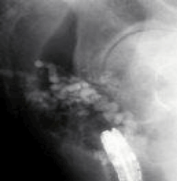
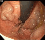
| Various medical treatments such as endoscopic treatments and interventional radiology have been used to control bleeding from rectal varices, however, there is no best treatment strategy for rectal varices. Surgical treatments, including portosystemic shunting, ligation, and under-running suturing have been performed [1]. In patients with poor condition, interventional radiologic techniques such as transjugular intrahepatic portosystemic shunts were successfully employed for rectal variceal bleeding as a non-operative treatment option [7-9]. Wang et al. found the usefulness of EIS in treating rectal variceal bleeding [10]. On the other hand, Levine et al. treated rectal varices initially with EIS, and 1 week later, EBL was performed on the remaining rectal varices as additional treatment [11]. EBL was introduced as a new method for treating esophageal varices, and it is reportedly both easier to perform and safer than EIS. Some reports revealed the usefulness of EBL in several cases as a treatment of rectal varices [12,13], and described EBL as a safe and effective therapy for rectal varices [13]. EBL may be effective as an initial treatment for rectal variceal bleeding, however, some investigators reported the easily recurrence of rectal varices after EBL [14,15]. Sato et al. performed EIS using 5% ethanolamine oleate with iopamidol (5%EOI), under fluoroscopy without complications, and they retrospectively evaluated the rates of recurrence of rectal varices after EIS or EBL [15]. The rates of recurrence of rectal varices tended to be more frequent with EBL than EIS and they mentioned that it is important to evaluate the hemodynamics of the rectal varices before EIS procedure to avoid severe complications such as vascular embolism, and 5%EOI of the sclerosant should be slowly injected under fluoroscopy, taking care to ensure that the agent does not flow into the systemic circulation during EIS. We show a case of rectal variceal patient, and present endoscopic findings before EIS (Figure 1a) and the fluoroscopy during EIS (Figure 1b). Shudo et al. successfully performed concurrent EBL and EIS treatment for a case of rectal varices [16]. |
| Percutaneous transhepatic obliteration (PTO) may be useful for large rectal varices. Sato et al. show a cirrhotic case with anal bleeding of large tortuous rectal varices [17]. Percutaneous transhepatic portography was performed by catheterization of the right anterior branch of the intrahepatic portal vein. Hepatofugal blood flow was observed in the superior rectal vein connected to the inferior mesenteric vein and the contrast medium passed into the rectal varices. Next, PTO was performed with injection of 5% ethanolamine oleate containing iopamidol and Histoacryl diluted with Lipiodol via the afferent veins of rectal varices in this case, and computed tomography revealed the successfully therapeutic effect in showing the accumulation of Lipiodol in rectal varices after PTO. A standard best treatment strategy for rectal varices has not been established. It is necessary to evaluate the suitable evidence-based treatment for rectal varices in larger numbers of patients with more investigations. |
| Conclusion |
| 1. Hemorrhage from rectal varices should be kept in mind in patients with portal hypertension presenting with lower gastrointestinal bleeding. |
| 2. Endoscopic procedures such as EIS and EBL are useful for rectal varices. PTO may be useful for large rectal varices. However, a best medical treatment strategy for rectal varices has not been established. It is necessary to evaluate the suitable evidencebased treatment for rectal varices in larger numbers of patients with more investigations. |
References
- Hosking SW, Smart HL, Johnson AG, Triger DR (1989) Anorectal varices, haemorrhoids, and portal hypertension. Lancet 1: 349-352.
- Wang TF, Lee FY, Tsai YT, Lee SD, Wang SS, et al. (1992) Relationship of portal pressure, anorectal varices and hemorrhoids in cirrhotic patients. J Hepatol 15: 170-173.
- Chawla Y, Dilawari JB (1991) Anorectal varices--their frequency in cirrhotic and non-cirrhotic portal hypertension. Gut 32: 309-311.
- McCormack TT, Bailey HR, Simms JM, Johnson AG (1984) Rectal varices are not piles. Br J Surg 71: 163.
- Johansen K, Bardin J, Orloff MJ (1980) Massive bleeding from hemorrhoidal varices in portal hypertension. JAMA 244: 2084-2085.
- Wilson SE, Stone RT, Christie JP, Passaro E Jr (1979) Massive lower gastrointestinal bleeding from intestinal varices. Arch Surg 114: 1158-1161.
- Katz JA, Rubin RA, Cope C, Holland G, Brass CA (1993) Recurrent bleeding from anorectal varices: successful treatment with a transjugular intrahepatic portosystemic shunt. Am J Gastroenterol 88: 1104-1107.
- Shibata D, Brophy DP, Gordon FD, Anastopoulos HT, Sentovich SM, et al. (1999) Transjugular intrahepatic portosystemic shunt for treatment of bleeding ectopic varices with portal hypertension. Dis Colon Rectum 42: 1581-1585.
- Fantin AC, Zala G, Risti B, Debatin JF, Schöpke W, et al. (1996) Bleeding anorectal varices: successful treatment with transjugular intrahepatic portosystemic shunting (TIPS). Gut 38: 932-935.
- Wang M, Desigan G, Dunn D (1985) Endoscopic sclerotherapy for bleeding rectal varices: a case report. Am J Gastroenterol 80: 779-780.
- Levine J, Tahiri A, Banerjee B (1993) Endoscopic ligation of bleeding rectal varices. Gastrointest Endosc 39: 188-190.
- Firoozi B, Gamagaris Z, Weinshel EH, Bini EJ (2002) Endoscopic band ligation of bleeding rectal varices. Dig Dis Sci 47: 1502-1505.
- Sato T, Yamazaki K, Toyota J, Karino Y, Ohmura T, et al (1999) Two Cases of Rectal Varices Treated by Endoscopic Variceal Ligation. Dig. Endosc 11: 66-69.
- Shudo R, Yazaki Y, Sakurai S, Uenishi H, Yamada H, et al (2000) Endoscopic Variceal Ligation of Bleeding Rectal Varices: A Case Report. Dig. Endosc 12: 366-368.
- Sato T, Yamazaki K, Toyota J, Karino Y, Ohmura T, et al. (2006) The value of the endoscopic therapies in the treatment of rectal varices: a retrospective comparison between injection sclerotherapy and band ligation. Hepatol Res 34: 250-255.
- Shudo R, Yazaki Y, Sakurai S, Uenishi H, Yamada H, et al (2001) Combined endoscopic variceal ligation and sclerotherapy for bleeding rectal varices associated with primary biliary cirrhosis: a case showing a long-lasting favorable response. Gastrointestinal Endosc 53:661-665.
- Sato T, Yamazaki K, Akaike J (2009) Clinical features of ectopic varices. JJPH 15:149-53.
Figures at a glance
 |
 |
| Figure 1 | Figure 2 |
Relevant Topics
- Constipation
- Digestive Enzymes
- Endoscopy
- Epigastric Pain
- Gall Bladder
- Gastric Cancer
- Gastrointestinal Bleeding
- Gastrointestinal Hormones
- Gastrointestinal Infections
- Gastrointestinal Inflammation
- Gastrointestinal Pathology
- Gastrointestinal Pharmacology
- Gastrointestinal Radiology
- Gastrointestinal Surgery
- Gastrointestinal Tuberculosis
- GIST Sarcoma
- Intestinal Blockage
- Pancreas
- Salivary Glands
- Stomach Bloating
- Stomach Cramps
- Stomach Disorders
- Stomach Ulcer
Recommended Journals
Article Tools
Article Usage
- Total views: 17924
- [From(publication date):
specialissue-2013 - Apr 04, 2025] - Breakdown by view type
- HTML page views : 13271
- PDF downloads : 4653
