Case Report Open Access
Skeletal Muscle Metastasis Secondary to Adenocarcinoma of Colon: A Case Report and Review of Literature
| Mutahir A Tunio1*, Mushabbab Al Asiri1, Khalid Riaz1, Wafa Al Shakwer2 and Muhannad Al Arifi3 | |
| 1Radiation Oncology, Comprehensive Cancer Center, King Fahad Medical City, Riyadh-59046, Saudi Arabia | |
| 2Histopathology, Comprehensive Cancer Center, King Fahad Medical City, Riyadh-59046, Saudi Arabia | |
| 3King Saud Bin Abdul Aziz University for Health Sciences, Riyadh 11345, Saudi Arabia | |
| Corresponding Author : | Mutahir A. Tunio Assistant Consultant Radiation Oncology Comprehensive Cancer Center King Fahad Medical City, Riyadh, Saudi Arabia E-mail: drmutahirtonio@hotmail.com |
| Received February 21, 2013; Accepted March 16, 2013; Published March 18, 2013 | |
| Citation: Tunio MA, AlAsiri M, Riaz K, AlShakwer W, AlArifi M (2013) Skeletal Muscle Metastasis Secondary to Adenocarcinoma of Colon: A Case Report and Review of Literature. J Gastroint Dig Syst S12:002. doi: 10.4172/2161-069X.S12-002 | |
| Copyright: © 2013 Tunio MA, et al. This is an open-access article distributed under the terms of the Creative Commons Attribution License, which permits unrestricted use, distribution, and reproduction in any medium, provided the original author and source are credited. | |
Visit for more related articles at Journal of Gastrointestinal & Digestive System
Abstract
Introduction: Colon adenocarcinoma frequently metastasizes to the liver, regional lymph nodes, lungs and peritoneum. However, metastasis to the skeletal muscles is extremely rare manifestation of colon adenocarcinoma. To date, only few cases have been reported in the literature. Skeletal muscle metastasis from colon adenocarcinoma usually remains asymptomatic or manifest as swelling and are associated with dismal prognosis. Case presentation: A 28 years old Saudi man known case of adenocarcinoma of transverse colon treated with extended hemi-colectomy and chemotherapy one year back, presented with abdominal wall swelling and right buttock swelling since 8 months. Physical examination revealed right gluteal mass of size 3×2 cm and abdominal wall mass of size 2×2cm. Rest of examination was unremarkable. Computed tomography-Positron emission tomography (CT-PET) showed 3×2 cm lobulated mass arising from gluteus maximus muscle and another mass in rectus abdominis muscle. Incisional biopsy confirmed the metastatic adenocarcinoma of colon. Patient subsequently underwent palliative radiotherapy followed by systemic chemotherapy. At time of publication, patient was alive with progressive disease. Conclusion: Skeletal muscles metastases are rare manifestation of adenocarcinoma of colon and searching for the primary focus in a patient with skeletal muscle metastasis, colon cancer should be considered as differential diagnosis.
| Keywords |
| Colon adenocarcinoma; skeletal muscle metastasis; Rare |
| Introduction |
| Colon cancer is less common in the Saudi Arabia than in its counterpart Gulf Cooperation Council (GCC) States and in the West, but the incidence is increasing, making this disease he second most common malignancy after breast cancer, ranking first among men and third among women between 1994 and 2004 [1]. In 2004, 647 new cases of colon cancer were diagnosed in the Saudi Arabia with median age of 60 years in men (range, 19-105 years) and 58 years in women (range 16- 100 years) [1].Unfortunately most colon cancers present at metastatic stages in our region, and are not amenable to upfront curative surgery. |
| While the liver and lung are primary targets for distant metastasis from colon cancer, the metastasis to other distant sites is rarely found. Metastasis from colon cancer to skeletal muscles is rarest manifestation, only few related case reports have been published in literature so far [2], and the prognosis is generally dismal with reported median survival from 5-12 months. |
| Here-in, we report a 28 years old Saudi man with skeletal muscle metastasis to gluteus maximus and rectus abdominis as manifestation of mucinous adenocarcinoma of transverse colon. |
| Case Presentation |
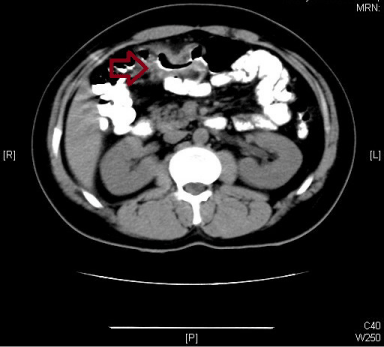
| A 28 years old Saudi male presented in our clinic on February 2011, with 5-month history of abdominal discomfort, altered bowl habits and off and on rectal bleeding and 3 kg weight loss. He had no history of any medical illness and smoking and there no family history of malignancy. On physical examination he was found emaciated, mild anemic with no palpable lymphadenopathy and visceromegaly. Colonoscopy showed mass at splenic flexure of transverse colon and biopsy showed moderately differentiated adenocarcinoma. Baseline carcino-embryonic antigen (CEA) level was 117 ng/ml and computed tomography (CT) of abdomen showed hepatic flexure of transverse colon mass however there were no pulmonary and liver metastasis (Figure 1). Then he underwent extended hemi-colectomy and ileostomy. Histopathology showed, mucinous moderately differentiated adenocarcinoma penetrating into muscularis propria, serosa and pericolic fat. Proximal and distal margins were found negative. Seven out of 13 retrieved lymph nodes were found metastatic and extra-nodal extension was seen in 2 lymph nodes. Multiple peritoneal nodules were also resected and biopsy was consistent with mucinous adenocarcinoma. His final stage was pT4N2bM0. Patient was planned for adjuvant chemotherapy but he lost to follow up. |
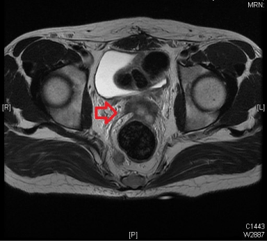
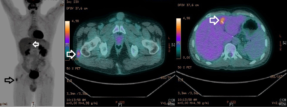
| In January 2012, he presented in clinic with abdominal pain and alerted bowel habits. On physical examination, there was hard nodule was felt at anterior abdominal wall just below the umbilicus and another nodule was found at right lateral thigh. Both nodules were mild tender; however rest of examination was unremarkable. Magnetic resonance imaging (MRI) showed multiple extensive peritoneal masses, the largest noted within the rectovaginal pouch of size 3×5.7 ×3.4 cm, invading the base of the urinary bladder, sigmoid, seminal vesicles as well as the distal part of the left ureter (Figure 2). Other peritoneal lesions noted within the right and left iliac fossa as well as the perirectal fossa, invading both ureters in the pelvic sidewalls and peritoneal metastasis on the surface of the liver near segment VI and VII. Positron emission tomography-CT (PET-CT) showed additional skeletal muscle metastasis of rectus abdominis muscle and right gluteus maximus (Figure 3). Incisional Biopsies of rectus abdominis and right gluteal muscle masses were consistent with metastatic adenocarcinoma (Figure 4). Patient was given FOLFOX-4 12 cycles after the JJ stenting and then was treated with radiation therapy for rectus abdominis and gluteal muscles metastases with 6MV photons with total dose 45 Gy in 25 fractions. Patient had progression of disease and he was started on second line chemotherapy FOLFIRI. Patient was alive at 12 months after discovery of skeletal muscle metastasis. |
| Discussion |
| Even though skeletal muscle constitutes about 40% of body weight, is the largest organ in the human body, but the skeletal muscle metastasis is a rare entity and differentiation between a primary soft tissue sarcoma and metastatic carcinoma is difficult without biopsy [3]. Most common malignancies metastasizing to skeletal muscles are lung, stomach and genitourinary tumors. Skeletal muscle metastases manifest as painful masses of size 2-12cm [4]. Metastasis from colon cancer to skeletal muscles is very rare manifestation, so far only eight related case reports have been published in literature. [5-12] (Table 1) |
| The possible mechanism of metastatic spread of adenocarcinoma of colon to the skeletal muscles could be by lymphatics, hematogenous route, and direct extension of primary disease and by manipulation during surgery [13]. In our patient, abdominal wall and gluteal muscle metastases could be due to surgical implantation. Having obtained clear lateral margins at the initial surgery, the metastases were likely to have been secondary to the seeding of exfoliated tumor cells during tumor mobilization [14]. |
| Skeletal muscle metastases are commonly thought to be associated with poor average survival because of underlying widespread disease, with an average of 5-12 months after diagnosis; it warrants aggressive surgical resection, radiotherapy and the use of systemic chemotherapy [15]. In our patient resection was not done because of wide spread peritoneal metastasis. |
| In conclusion, we have reported a case of colon cancer with gluteus maximus and rectus abdominis muscle metastasis, which is extremely rare. CT- PET can be helpful to localize the site for metastatic deposits and immunostaing whenever possible shall be incorporated to reach the final diagnosis and for prompt treatment. |
References
- Saudi Cancer Registry (SCR) Saudi Arabia Cancer Incidence and Survival Report 2007.
- Plaza JA, Perez-Montiel D, Mayerson J, Morrison C, Suster S (2008) Metastases to soft tissue: a review of 118 cases over a 30-year period. Cancer 112: 193-203.
- Seely S (1980) Possible reasons for the high resistance of muscle to cancer. Med Hypotheses 6: 133-137.
- Tuoheti Y, Okada K, Osanai T, Nishida J, Ehara S, et al. (2004) Skeletal muscle metastases of carcinoma: a clinicopathological study of 12 cases. Jpn J Clin Oncol 34: 210-214.
- Takada J, Watanabe K, Kuraya D, Kina M, Hayashi S, et al. (2011) An example of metastasis to the iliopsoas muscle from sigmoid colon cancer. Gan To Kagaku Ryoho 38: 2294-2297.
- Naik VR, Jaafar H, Mutum SS (2005) Heterotopic ossification in skeletal muscle metastasis from colonic adenocarcinoma--a case report. Malays J Pathol 27: 119-121.
- Burgueño Montañés C, López Roger R (2002) Exophthalmos secondary to a lateral rectus muscle metastasis. Arch Soc Esp Oftalmol 77: 507-510.
- Hasegawa S, Sakurai Y, Imazu H, Matsubara T, Ochiai M, et al. (2000) Metastasis to the forearm skeletal muscle from an adenocarcinoma of the colon: report of a case. Surg Today 30: 1118-1123.
- Homan HH, Mühlberger T, Kuhnen C, Steinau HU (2000) Intramuscular extremity metastasis of adenocarcinoma of the colon. Chirurg 71: 1392-1394.
- Bonjer HJ, Lange JF, Jansen A, Schouten WR, Jakimowicz J, et al. (1997) Abdominal wall metastasis following surgical removal of colorectal carcinomas. Ned Tijdschr Geneeskd 141: 1868-1870.
- Jacquet P, Averbach AM, Jacquet N (1995) Abdominal wall metastasis and peritoneal carcinomatosis after laparoscopic-assisted colectomy for colon cancer. Eur J Surg Oncol 21: 568-570.
- Laurence AE, Murray AJ (1970) Metastasis in skeletal muscle secondary to carcinoma of the colon--presentation of two cases. Br J Surg 57: 529-530.
- De Friend DJ, Kramer E, Prescott R, Corson J, Gallagher P (1992) Cutaneous perianal recurrence of cancer after anterior resection using the EEA stapling device. Ann R Coll Surg Engl 74: 142-143.
- Tunio MA, Hashmi A, Mubarik M (2010) Skin and Skeletal Muscle Metastasis of adenocarcinoma of rectum: an unusual manifestation. J of Surg Pak 15:203-205
- Glockner JF, White LM, Sundaram M, McDonald DJ (2000) Unsuspected metastases presenting as solitary soft tissue lesions: a fourteen-year review. Skeletal Radiol 29: 270-274.
Tables and Figures at a glance
| Table 1 |
Figures at a glance
 |
 |
 |
 |
| Figure 1 | Figure 2 | Figure 3 | Figure 4 |
Relevant Topics
- Constipation
- Digestive Enzymes
- Endoscopy
- Epigastric Pain
- Gall Bladder
- Gastric Cancer
- Gastrointestinal Bleeding
- Gastrointestinal Hormones
- Gastrointestinal Infections
- Gastrointestinal Inflammation
- Gastrointestinal Pathology
- Gastrointestinal Pharmacology
- Gastrointestinal Radiology
- Gastrointestinal Surgery
- Gastrointestinal Tuberculosis
- GIST Sarcoma
- Intestinal Blockage
- Pancreas
- Salivary Glands
- Stomach Bloating
- Stomach Cramps
- Stomach Disorders
- Stomach Ulcer
Recommended Journals
Article Tools
Article Usage
- Total views: 14084
- [From(publication date):
specialissue-2013 - Nov 21, 2024] - Breakdown by view type
- HTML page views : 9678
- PDF downloads : 4406
