Secondary Haemophagocytosis in a Patient with Ewing's Sarcoma
Received: 17-Sep-2013 / Accepted Date: 22-Nov-2013 / Published Date: 25-Nov-2013 DOI: 10.4172/2161-0681.1000151
Abstract
Secondary haemophagocytosis in malignant tumours have been reported in bone marrow. However we present engulfment of malignant apoptotic Ewing’s sarcoma cells by macrophages in the bone marrow. A 24 year old Pakistani male patient presented with complains of backache. After imaging, a laminectomy was performed the biopsy results of which were consistent with Ewing’s sarcoma. Subsequently for staging, bone marrow and trephine was done. The bone marrow aspirate showed markedly increased haemophagocytic activity with histiocytes engulfing malignant apoptotic Ewing’s sarcoma cells. The presence of histiocytes engulfing apoptotic Ewing’s sarcoma cells was of particular interest. Similar findings have rarely been reported.
Keywords: Bone marrow; Haemophagocytosis; Ewing’s sarcoma
309444Introduction
In children and young adults, Ewing’s sarcoma is the second most common type of primary bone malignancy. Secondary haemophagocytosis in bone marrow aspirate is a common finding previously described as malignant histiocytosis. Previously Linn et al. [1] has reported n=10 cases of malignant histiocytosis mostly seen in high grade lymphomas. It has a poor prognosis and is refractory to treatment modalities [2]. Our patient showed presence of secondary haemophagocytosis in bone marrow with a primary diagnosis of Ewing’s sarcoma. The bone marrow aspirate showed multiple macrophages engulfing malignant apoptotic Ewing’s sarcoma cells. Secondary haemophagocytic in Ewing’s sarcoma has rarely been reported.
Case Presentation
A 24 year old Pakistani male presented with complains of backache for three months. He had a road traffic accident three months back and was correlating the pain with the incident. Finally, after no relief with painkillers and additional symptoms of weight loss, he visited his physician and was evaluated. Complete blood counts were done which showed Hb: 12.2 g/dl, WBC: 7.4 × 10⁹/L and Platelets: 265 × 109/L. Peripheral blood film revealed normocytic, normochromic red blood cells, left shifted neutrophils with toxic granulations and normal platelets. Because of the persistent backache he was then referred to Neurology. MRI imaging was performed initially which displayed fracture of 8th vertebral body with edema. Discernible retropulsion of posterior fragment compressing ventral thecal sac with slightly effaced bilateral neuroforamina and well preserved nerve roots was also seen. Subsequently he underwent laminectomy. Post surgery MRI imaging was repeated. The results were reported as multilevel lytic lesions involving mid dorsal vertebrae (D5-D7) with marrow infiltration. There was also a soft tissue mass having prevertebral and intraspinal components.
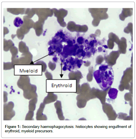
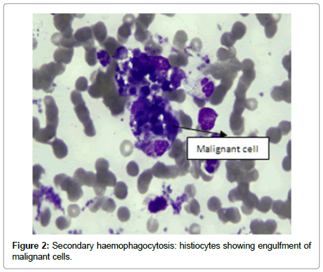
The laminectomy sample was sent for histopathology which was reported as Ewing’s sarcoma. As a staging procedure, bone marrow and trephine was done. Bone marrow sample showed presence of normal haematopoietic precursors along with infiltration of malignant tumour cells. These were large atypical mononuclear cells with intense basophilic cytoplasm and vacuolations. The nuclei of these cells revealed open chromatin. The bone marrow sample also showed presence of numerous histiocytes which had engulfed erythroid, myeloid precursors and megakaryocytes (Figures 1 and 2). Few of the histiocytes had engulfed malignant apoptotic cells. Bone trephine revealed normal cellularity for age, all haematopoietic precursors and a single fibrosed area which did not show positivity for CD99 due to underlying necrosis.
Conclusion
Bone marrow aspirate showed presence of malignant apoptotic Ewing’s sarcoma cells being engulfed by macrophages. These findings have rarely been reported.
References
- Linn YC, Tien SL, Lim LC, Lee LH, Teoh G, et al. (1995) Haemophagocytosis in bone marrow aspirate-a review of the clinical course of 10 cases. Acta Haematol 94: 182-191.
- Emmenegger U, Reimers A, Frey U, Fux Ch, Bihl F, et al. (2002) Reactive macrophage activation syndrome: a simple screening strategy and its potential in early treatment initiation. Swiss Med Wkly 132: 230-236.
Citation: Ali N, Mansoori H (2013) Secondary Haemophagocytosis in a Patient with Ewing’s Sarcoma. J Clin Exp Pathol 4:151. DOI: 10.4172/2161-0681.1000151
Copyright: © 2013 Ali N, et al. This is an open-access article distributed under the terms of the Creative Commons Attribution License, which permits unrestricted use, distribution, and reproduction in any medium, provided the original author and source are credited.
Share This Article
Recommended Journals
Open Access Journals
Article Tools
Article Usage
- Total views: 16050
- [From(publication date): 1-2014 - Apr 05, 2025]
- Breakdown by view type
- HTML page views: 11345
- PDF downloads: 4705


