| Research Article |
Open Access |
|
| Giuliana Pace*, Marco Caruso, Vincenzo Sucato, Giuseppe Riccardo Tona, Angelo Quagliana, Debora Cangemi, Deborha Maniglia, Fiorella Sutera, Giovanna Evola, Salvatore Evola and Salvatore Novo |
| Department of Internal Medicine, Cardiovascular and NefroUrologic Disease of University of Palermo, Italy |
| *Corresponding authors: |
Giuliana Pace
Department of Internal Medicine
Cardiovascular and NefroUrologic Disease of University of Palermo
Via Del Vespro, 129, 90100 Palermo, Italy
Tel: 0916552986
Fax: 0916554301
E-mail: giuliana.pace@libero.it |
|
| |
| Received November 23, 2011; Published October 30, 2012 |
| |
| Citation: Pace G, Caruso M, Sucato V, Tona GR, Quagliana A, et al. (2012) Clinical Appropriateness of Coronary Angiography. 1:419. doi:10.4172/scientificreports.419 |
| |
| Copyright: © 2012 Pace G, et al. This is an open-access article distributed under the terms of the Creative Commons Attribution License, which permits unrestricted use, distribution, and reproduction in any medium, provided the original author and source are credited. |
| |
| Abstract |
| |
| Background: The study evaluates the appropriateness of coronary angiography and the agreement between the used method and the presence of coronary artery disease by the indications proposed from American College of Cardiology/American Heart Association (1999). |
| |
| Method: The guidelines allow us to associate to Class I and IIa the judgment of appropriateness, to the Class IIb of uncertainty; to Class III of inappropriateness. |
| |
| Result: On 761 coronary angiography 76.74% were appropriate, 23.13% unsuitable, 0.13% uncertain. The group with the greater value of appropriateness is that one with unstable angina (97.9% appropriate); that one with the lower value is the group with non-specific symptomatology (26.7% appropriate). |
| |
| Conclusion: Considering the false positives, it is important the rate of the greater sensibility and the lower specificity of the not invasive tests carried before coronary angiography, as well as, the probable presence of microcircle disease. Among the false negatives, we must considered the number of patients with effective coronary artery disease which has “jumped” the intermediate stage of the not invasive diagnostic process, before the coronary angiography, but have obtained the same final benefit. |
| |
| Keywords |
| |
| Atherosclerosis; Acute coronary syndromes; Percutaneous coronary intervention; Appropriateness of coronary angiography examination |
| |
| Abbreviations |
| |
| ACC/AHA: American College of Cardiology/ American Heart Association; FP: False Positives; TP: True Positives; MI: Myocardical Infarction; PCI: Percutaneous Coronary Intervention |
| |
| Introduction |
| |
| According to the definition of Brook et al. [1], a procedure is considered "appropriate" if the expected benefit in terms of health is greater than complications it entails with a sufficiently wide margin. However, the concept of appropriateness is useful if added to the one of "necessity": the procedure must be done because otherwise the patient could have a damage [2,3]. For this purpose, the method used is that developed by the RAND Corporation in collaboration with the University of California Los Angeles Health Service Utilization Study [ 4]. Another popular method consist of comparing the directions to make angiography of the analized population with those described in the guidelines, in general that of American Heart Association/American College of Cardiology and the European Society of Cardiology [5]. The criteria did not show only temporal variability, but also geographical [ 6-10], in fact, those adopted in the United States tended to be wider than in Europe and Canada [11-13]. To evaluate the appropriateness of a diagnostic examination, a diagnostic procedure to be applied to all patients, should be established a priori, in order to submit a smaller number of patients at risk. |
| |
| Methods |
| |
| The purpose of this study is to evaluate the appropriateness of the coronary angiography in order to verify the correlation between the method used, the real presence of disease and its severity. To this end, a retrospective analysis was performed. We considered 761 patients, of whom 558 men and 203 women aged between 31 and 92 years; were analyzed: diagnosis of acceptance, the major cardiovascular risk factors, noninvasive functional assessment (ergometric test, stress echo, perfusion myocardial scintigraphy, and echocardiogram), report of CVG. Patients were divided into nine groups carefully following the criteria proposed by the guidelines ACC/AHA for the indication for coronary angiography: |
| |
• Stable angina/Asymptomatic
• Unstable angina
• Post-ischemic revascularization
• MI stratification post-MI
• Chest pain of undetermined origin
• Heart failure • Valvulopathy
• Nonspecific symptoms (asthenia, syncope, dyspnea)
• Pre-surgery for non-cardiac disease |
| |
| Analogous to the study of Rubboli et al. [14], the coronary angiographies performed in patients whose clinical condition and the previously diagnostic process which resided in Class I, were considered appropriate; the same is for the Class IIa. Class III belongs to all those conditions in which the indication for coronary angiography involves more risks than benefits, for which, considered inappropriate. |
| |
| Results |
| |
| Patients were divided into nine groups and it was observed that the greatest numbers of coronary angiographies were performed in patients with MI, followed by patients with unstable angina and stable angina/asymptomatic (Table 1). The 36.3% of all appropriate coronary angiographies were performed in patients with MI. The highest rate of inappropriateness is reached among patients with nonspecific symptoms (73.33%) followed by those with chest pain of undetermined origin (63.33%), while the lowest rate belongs to the category of patients with unstable angina (2.05%). During the evaluation it was considered the weight of each cardiovascular risk factor in the outcome of the study. As shown in figure 1, the rate of appropriateness is constant, for any number of risk factors, between 65% and 80%. The appropriateness of the values varies from 60% for the group 31-35 years to values of 100% for that of 91-95 years, but since this group consists of only one patient, the value is distorted: eliminating this group, however, the value of appropriateness, remains high and steady, fluctuating between 60% (31-35 years) and 90% (81-85 years) with a mean of 75% (Figure 2). |
| |
|
|
Table 1: Distribution per categories of appropriate, inappropriate, dubious coronary angiography. |
|
| |
|
|
Figure 1: Distribution of the coronary angiography appropriateness stratified by number of risk factors (rates). |
|
| |
|
|
Figure 2: Distribution of the appropriateness stratified by age groups (rates). |
|
| |
| Discussion |
| |
| The analysis of the obtained data shows that On 761 patients considered, 584 (76.74%) have made appropriate coronary angiography, 176 (23.13%) have performed inappropriate coronary angiography and only 1 coronary angiography was uncertain indication (0.13%) (Table 2). The total number of coronary angiographies (761), 584 (76.74%) showed a hemodynamically significant stenosis of coronary tree which interesting one or more arterial branches (Figure 3). |
| |
|
|
Table 2: Overall assessment of the angiographies. |
|
| |
| |
|
|
Figure 3: Result of angiographies carried out on the whole population analyzed. |
|
| |
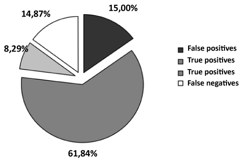
| The population analyzed was performed 263 angioplasties and 192 have been put to surgical revascularization. Table 3 shows the distribution of appropriate and inappropriate coronary angiographies on the basis of the angiographic result. Of 177 coronary angiographies that showed coronary free, 114 were appropriate (64.41%); of 584 coronary angiographies that showed coronary stenosis, 470 were appropriate (80.48%). Considering all of the appropriate angiographies (n=584) it is clear that 470 showed hemodynamically significant coronary stenosis (80.48% of all appropriate coronary angiography): this group shows the true positives, i.e. those patients in whom the diagnostic procedure was correct, then the indication was appropriate, and have coronary artery disease. These patients represent the largest group being 62% of total patients. |
| |
|
|
Table 3: Correlation between angiographic results and appropriateness; absolute number of False Positives (FP), True Positive (TP), True Negative (TN), False Negative (FN). |
|
| |
| The group, in which there were made coronary angiography appropriate for indication, but no evidence of coronary artery disease, represents false positives, i.e. those patients in whom the correct diagnostic procedure does not correlate to the anatomic description. These are 114 patients, 20% of all patients with appropriate coronary angiography. To this time we have tried to explain the presence of this last group, and we made more assumptions. The first, and also the most obvious, is that the information provided by ACC/AHA should be revised in the light of the methods with greater sensitivity and specificity we have, and of new knowledge in theme of atherosclerosis; the second reason may relate to the spectrum of specificity, probably not wide enough, of non-invasive methods and in this case of ergometric test that most of our patients performed as non-invasive screening method; may also be the high sensitivity of a method such as myocardial scintigraphy, used in 24 of 114 patients FP (i.e. 21%), could identify a step ahead of the ischemic cascade, as well as the alteration of coronary microcirculation. |
| |
| The third group consists of patients with inappropriate coronary angiography and coronaries free which are the true negatives, i.e. those patients in whom it was necessary to stop the diagnostic process before the coronary angiography as the previous non-invasive investigations had proved negative, or in the presence of an equivocal ergometric test, needed to proceed to iterate through another non-invasive method before coronary angiography. These patients, however, represent the smallest group being only 8% of the total. Finally, considering the group of false negatives, i.e. 113 patients (i.e. 13% of the total and 64.20% of all inappropriate coronary angiography) with inappropriate investigation but diseased coronary arteries, it was clear that these subjects has probably stopped the diagnostic process prematurely as they should make other non-invasive diagnostic methods before coronary angiography. |
| |
| Conclusions |
| |
| The data we obtained, ultimately, are consistent with those reported in literature: patients with appropriate investigations are 76.74%, 23.13% on the inappropriate, uncertain 0.13%. Among appropriates the most are true positives: this indicates proper adherence to the diagnostic protocol. Among the inappropriate group, the less represented is that of true negatives. The other two groups of so-called "false" merit particular considerations. False positives could actually be reduced by 21% considering that probably the low specificity of scintigraphy implies a detailed reason: in fact the scintigraphy FP considered by angiographic anatomy standard, are actually the TP comparing to a pathophysiological standard like coronary reserves or endothelial function (Figure 4). |
| |
|
|
Figure 4: Distribution of the population subjected to diagnostic process. |
|
| |
| As far as the false negative (n=113) is concerned, we wondered, what could be the therapeutic fate of these subjects. Table 4 summarizes the therapy of these patients based on angiographic result: 44 of them were considered worthy of surgical revascularization, 45 patients were treated with coronary angioplasty during the same session, and 24 were discharged with medical therapy. |
| |
|
|
Table 4: Therapeutic indications in patients with inappropriate coronary angiography and coronary disease. |
|
| |
| |
| References |
| |
- Brook RH, Chassin MR, Fink A, Solomon DH, Kosecoff J, et al. (1986) A method for the detailed assessment of the appropriateness of medical technologies. Int J Technol Assess Health Care 2: 53-63.
- Kahan JP, Bernstein SJ, Leape LL, Hilborne LH, Park RE, et al. (1994) Measuring the necessity of medical procedures. Med Care 32: 357-365.
- Guadagnoli E, Landrum MB, Peterson EA, Gahart MT, Ryan TJ, et al. (2000) Appropriateness of coronary angiography after myocardial infarction among Medicare beneficiaries. Managed care versus fee for service. N Engl J Med 343: 1460-1466.
- Fitch K, Bernstein SJ, Aguilar MD, Burnandet B, LaCalle JR, et al. (2001) The RAND/ UCLA Appropriateness Method User’s Manual. RAND News and Publications MR 1269.
- Scanlon PJ, Faxon DP, Audet AM, Carabello B, Dehmer GJ, et al. (1999) ACC/AHA guidelines for coronary angiography: executive summary and recommendations. A report of the American College of Cardiology/American Heart Association Task Force on Practice Guidelines (Committee on Coronary Angiography) developed in collaboration with the Society for Cardiac Angiography and Interventions. Circulation 99: 2345-2357.
- Bressan M, Zanchetta M, Michieletto F, Pedrocco A, Zoppo F, et al. (1998) [Coronary angiography in two defined populations: Padua and Citadella]. G Ital Cardiol 28: 274-280.
- Bengtson A, Herlitz J, Karlsson T, Brandrup-Wognsen G, Hjalmarson A (1994) The appropriateness of performing coronary angiography and coronary artery revascularization in a Swedish population. JAMA 271: 1260-1265.
- Bernstein SJ, Hilborne LH, Leape LL, Fiske ME, Park RE, et al. (1993) The appropriateness of use of coronary angiography in New York State. JAMA 269: 766-769.
- Mozes B, Shabtai E (1994) The appropriateness of performing coronary angiography in two major teaching hospitals in Israel. Int J Qual Health Care 6: 245-249.
- Bressan M, Stritoni P, Razzolini R, Favaretti C, Bianchi A, et al. (1993) [Coronary angiography in a defined population: a pilot study of the residents of Padua]. Cardiologia 38: 225-229.
- Bernstein SJ, Kosecoff J, Gray D, Hampton JR, Brook RH (1993) The appropriateness of the use of cardiovascular procedures. British versus U.S. perspectives. Int J Technol Assess Health Care 9: 3-10.
- McGlynn EA, Naylor CD, Anderson GM, Leape LL, Park RE, et al. (1994) Comparison of the appropriateness of coronary angiography and coronary artery bypass graft surgery between Canada and New York State. JAMA 272: 934-940.
- Brook RH, Kosecoff JB, Park RE, Chassin MR, Winslow CM, et al. (1988) Diagnosis and treatment of coronary disease: comparison of doctors’ attitudes in the USA and the UK. Lancet 1: 750-753.
- Rubboli A, La Vecchia L, Casella G, Sangiorgio P, Bracchetti D (2001) Appropriateness of the use of coronary angiography in a population of patients with ischemic heart disease. Ital Heart J 2: 696-701.
|
| |
| |


