| Research Article |
Open Access |
|
| Wenzhong Zhang* |
| Department of Toxicological Assessment, China National Center for Food Safety Risk Assessment, China |
| *Corresponding author: |
Wenzhong Zhang
Department of Toxicological Assessment
China National Center for Food Safety Risk Assessment
Room 307, Panjiayuan Nanli 7 Hao, Chaoyang District
Beijing City, 100021, China
E-mail: zhangwz2002@sina.com |
|
| |
| Received July 04, 2012; Published October 29, 2012 |
| |
| Citation: Zhang W (2012) Nuclei DNA Slide: A Novel Method to Measure Biological Effects of Radiation Damage. 1:393. doi:10.4172/scientificreports.393 |
| |
| Copyright: © 2012 Zhang W. This is an open-access article distributed under the terms of the Creative Commons Attribution License, which permits unrestricted use, distribution, and reproduction in any medium, provided the original author and source are credited. |
| |
| Abstract |
| |
| Purpose: Although there are many physical and chemical radiation detectors, biological effects of radiation damage are hard to measure. So we aim to use a novel in vitro method to measure biological effects of radiation. |
| |
| Materials and methods: Acellular DNA, nuclei DNA fixed on slides, is used in our experiment. The nuclei DNA slides used as a model are radiated for 0, 5, 25 and 45 min with Ultraviolet (UV) lamp. The DNA damage of nuclei DNA slides was detected with comet assay, and the pictures of comet assay were measured by comet auto-analysis software- Comet A1.0, and Differences among groups are compared with SPSS 11.0. |
| |
| Results: When nuclei DNA slides are radiated with UV, the DNA damage is significantly, also there is timeresponse relationship between UV and DNA damage. |
| |
| Conclusions: The nuclei DNA slides can be used to measure biological effects of radiation, and the method is sensitive and specific to detect biological effects of radiation. |
| |
| Keywords |
| |
| Nuclei DNA slide, Biological effect, Radiation damage, Comet A1.0 |
| |
| Introduction |
| |
| There are many methods that can be used to detect and quantify radiation based on DNA damage, such as using cells and microorganism as models, Cadet’s et al. [1] study indicated that ultraviolet radiation damaged cellular DNA. The models of cells also delivered dose-response and time-response relationship the increase of Single Strand Break (SSB) and Double Strand Break (DSB) lesions in the DNA molecule obtained in the in vitro radiations [2]. It is reported that purified DNA and T4 endonuclease V was used as a gold standard for detection on UV damage to DNA [3]. Purified DNA molecules has been extensively used to study radiation during the last decades , the results obtained from such in vitro experiments are consistent with those from radiation of cells and tissues [4-6]. |
| |
| The methods above may involve DNA degradation, processing, staining, and labeling procedures, by which may obscure or even alter the DNA damage [7]. Also, purified DNA method was complex and difficult to carry out. So we address a simple in vitro model, full nucleus DNA slide to test radiation, which was simple to carry out and easy to standard. |
| |
| Materials and Methods |
| |
| Cell culture and preparation |
| |
| GC-56 cells (human gastric cancer cells) are easy to get in lab; the cells are used in the study. GC-56 cells are cultured in flasks in RPMI 1640 (GIBICO, USA), supplemented by 10% heat-inactivated fetal bovine serum (Gibco, USA) and 15 mg/ml Penicillin/Streptomycin (Sigma Aldrich, USA) mixture. Cells are maintained at 37°C in 5% CO 2 atmosphere (Thermo, Germany). Preparation of cells, to be brief, is to gather exponentially growing cells and add 0.25% Trypsin in PBS and to centrifuge them. Pellets are re suspended in 1% (wt/vol) lowmelting- point agarose (Bio-Rad Lab, USA); and concentration of cells is adjusted to 1 × 10⁶ cells/ml. |
| |
| Full nuclei DNA slides preparation |
| |
| Cells mixed with 1% (wt/vol) low-melting-point agarose (Bio- Rad, USA), are concentration-adjusted to 1 × 10⁶ cells/ml, and are sandwiched between a lower layer of 1% (wt/vol) normal-meltingpoint agarose (Bio-Rad, USA) and an upper layer of 1% (wt/vol) lowmelting- point agarose on slides. The slides are immersed in cell lysis solution (BIO-LAB, CHINA) for 2 hours at 4°C. Then, the full nuclei DNA slides are prepared, which can be stored at 4°C in saturated humidity for 2 months. |
| |
| UV radiation procedure |
| |
| For UVA radiation, three UV lamps with an emission wavelength peak at 365 nm were used to ra¬diate the nuclei DNA slides. The slides were dunked in buffer on ice and placed below the lamps about 20 cm, exposed for 0, 5, 25 and 45 min, there were two slides in every time point. |
| |
| DNA damage determination by comet assay |
| |
| DNA damage is quantified by the alkaline Comet Assay [8]. Slides are washed and incubated in ice-cold alkaline unwinding buffer (300 mM NaOH, 1mM EDTA Na2.2H2O, pH>13) for 30 min. After 20 min of electrophoresis at 25 V and 300mA, nuclei are stained with 0.03% (v/v) GELRED (BIO-LAB, CHINA) for 3 min. The specialized equipment for comet electrophoresis is used (BIO-LAB, CHINA).In each group, digital image of 200 cells (100 cells each slide) are randomly captured for analysis by Comet A1.0 software (BIO-LAB, CHINA). The parameters of DNA tail percentage, tail length, olive tail moment, length ratio of tail/head are used in analysis (Graph 1). |
| |
|
|
| |
| Statistical analysis |
| |
| Data are expressed as average ± SD (Standard Deviation). Differences among the groups are analyzed by ANOVA, for the inequality of the variance the data were confirmed, they are further analyzed by Games-Howell. Results are considered significantly different only P values <0.05. |
| |
| Results |
| |
| Nuclei DNA alterations on radiated slides |
| |
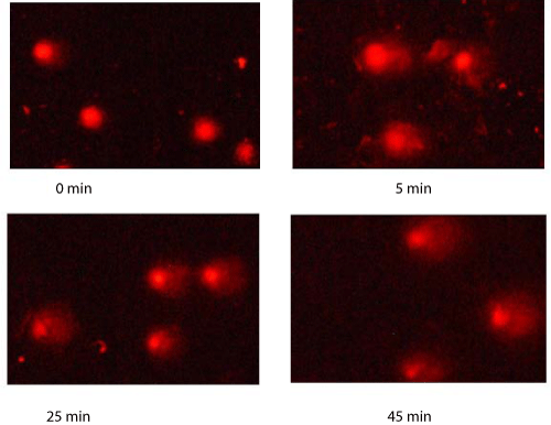
| To investigate the induced DNA damage with radiated slides, we analyze pictures of the comet assay. As mentioned before, DNA slides are exposed 0 min, 5 min, 25 min and 45 min under UV. The pictures show significant differences at time point. Figure 1 shows four comet assay images of nuclei DNA after UV radiation at 0 min, 5 min, 25 min and 45 min, respectively. With increasing exposure time, increasing length and DNA percent of comet tail are observed. |
| |
| Time-response relationship of the irradiated slides |
| |
| |
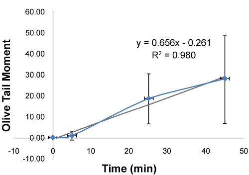
| The parameters of tail length, olive tail moment, tail DNA percent, and length ratio of tail/head are found to be treatment-dependent in this sequence: 45 min> 25 min> 5min> 0 min, as shown in table 1. Also there is time-response relationship between radiation and DNA damage, olive tail moment shows linear correlation, and there is significant time-response relationship between radiation and DNA damage, as shown in figure 1. |
| |
|
|
Table 1: Comet A Analyzed Results of Comet Pictures. |
|
| |
|
|
Figure 1: Comet Picture on Radiated DNA Slides. |
|
| |
| Discussion |
| |
| Acellular DNA is the popular method to study the DNA damage caused by radiation in vitro. A lots of reports are about research of plasmid DNA [6,8,9]. But the results analysis of plasmid DNA need to be scanned by CP-Research microscope and the test is very complex to carry out. Our method also uses a cellular DNA. Although the mechanism of two methods are almost similar, compare with other acellular, DNA slides have more benefits. Firstly, nuclei DNA slide method is simple and easy to carry out, all labs can use the same source commercial nuclei DNA slides; secondly nuclei DNA slides were easy to detect radiation, the nuclei DNA is fixed on the slides and can be stored for a long time before usage; thirdly, the nuclei DNA slides might be more sensitive than plasmid DNA, for nuclei DNA is long and centralized, and can carry out acellular comet assay [9]. |
| |
| Our study shows that the shape of the nuclei DNA changed significently after radiation, and the damage for 5 min’s radiation of UV could be observed. There were two possible reasons about shape changes of nuclei DNA, the first was that radiation increased the DNA strands break, and short strands easy diffuse; the second was that short strand’s electrophoresis speed was faster than longer strands. The DNA damage was also related with radiation intensity and time, 5 min UV radiation can be detected, and it’s indicated that the method was sensitive enough. Due to those benefits, nuclei DNA slides can be used to measure biological effects of radiation and anti-radiation in the future. |
| |
| In the study we use comet assay to analyze the nuclei DNA slides, it is considered that maybe there are other methods better than comet assay, for further research, we will combine and compare with different methods to measure the DNA damage on nuclei DNA slides. |
| |
| Conclusion |
| |
| In summary, nuclei DNA slides can be used to measure biological effects of radiation. For the nuclei DNA slides can be analyzed by acellular comet assay which is used widely, so the method of nuclei DNA slide to measure biological effects of radiation is simple, easy, cheap, sensitive, and specific, can be used broadly in the future. |
| |
| Acknowledgments |
| |
| This study is supported by the China CDC grant (grant No. 2011A205). None of the authors has financial interests related to this paper. None of the authors have financial interests related to this study. The authors would give special thanks to the Department of Food Toxicological Evaluation in the Institute of Nutrition and Food Safety, China CDC, for providing help in the study. |
| |
| |
| References |
| |
- Cadet J, Sage E, Douki T (2005) Ultraviolet radiation-mediated damage to cellular DNA. Mutat Res 571: 3-17.
- Goodhead DT, Thacker J, Cox R (1993) Weiss Lecture. Effects of radiations of different qualities on cells: molecular mechanisms of damage and repair. Int J Radiat Biol 63: 543-556.
- Douki T, Reynaud-Angelin A, Cadet J, Sage E (2003) Bipyrimidine photoproducts rather than oxidative lesions are the main type of DNA damage involved in the genotoxic effect of solar UVA radiation. Biochemistry 42: 9221-9226.
- Milligan JR, Arnold AD, Ward JF (1992) The effect of superhelical density on the yield of single-strand breaks in gamma-irradiated plasmid DNA. Radiat Res 132: 69-73.
- Leloup C, Garty G, Assaf G, Cristovão A, Breskin A, et al. (2005) Evaluation of lesion clustering in irradiated plasmid DNA. Int J Radiat Biol 81: 41-54.
- Milian FM, Gouveia AN, Gual MR, Echeimberg JO, Arruda-Neto JD, et al. (2007) In vitro effects of gamma radiation from 60Co and 137Cs on plasmid DNA. J Biol Phys 33: 155-160.
- Jiang Y, Rabbi M, Kim M, Ke C, Lee W, et al. (2009) UVA generates pyrimidine dimers in DNA directly. Biophys J 96: 1151-1158.
- Vasquez MZ (2010) Combining the in vivo comet and micronucleus assays: a practical approach to genotoxicity testing and data interpretation. Mutagenesis 25: 187-199.
- Kawaguchi S, Nakamura T, Honda G, Yokohama N, Sasaki Y (2008) Detection of DNA single strand breaks induced by chemicalmutagens using the acellular Comet assay. Genes and Environment, 30: 77-88.
|
| |
| |


