|
|
| Fischer BE*, Yates EW and Bhutta MA |
| Department of Trauma and Orthopaedics, Royal Bolton Hospital, Minerva Road, Farnworth, Bolton, UK |
| *Corresponding authors: |
Fischer BE
ST4, Trauma and Orthopaedics
Royal Bolton Hospital
Minerva Road, Farnworth
BL4 0JR, Bolton, UK
E-mail: benfischer77@gmail.com |
|
| |
| Received December 14, 2011; Published October 27, 2012 |
| |
| Citation: Fischer BE, Yates EW, Bhutta MA (2012) A Novel Approach to Fixing Medial Malleolus Fractures in the Presence of an Intramedullary Tibial Nail. 1:389. doi:10.4172/scientificreports.389 |
| |
| Copyright: © 2012 Fischer BE, et al. This is an open-access article distributed under the terms of the Creative Commons Attribution License, which permits unrestricted use, distribution, and reproduction in any medium, provided the original author and source are credited. |
| |
| Abstract |
| |
| A medial malleolus fracture can be difficult to stabilise when an ipsilateral diaphyseal fracture of the tibia has been treated with an intramedullary device. We describe a simple, reproducible technique using a “figure-of-8” tension band wire fixation, where the proximal hold for the tension band is provided by the distal locking bolt of the intramedullary device. |
| |
| Keywords |
| |
| Distal tibial metaphysis fracture; Medial malleolus fracture; Intramedullary device; Tension band wire |
| |
| Introduction |
| |
| Extra-articular fractures of the distal tibia are related to high-energy trauma, compressive force, direct bending or low-energy rotational force [1]. They are more common in men and are typically associated with road traffic collisions, falls from height and twisting injuries [ 2]. Fractures of the distal tibial metaphysis are thought to represent approximately 15% of all fractures of the distal third of the tibia. |
| |
| This fracture pattern has been defined and classified. A concomitant lateral malleolus fracture is often present and has been reported in up to 80% of cases. Associated posterior malleolus fractures and spiral fractures of the tibia have also been described in 0.6% to 25% of cases. A higher incidence of associated fractures is reported in more recent articles and this is possibly due to improvements in image clarity and the use of other imaging modalities including computed tomography (CT). |
| |
| The nature and incidence of combined ipsilateral medial malleolus and distal tibial metaphyseal fractures have not been defined in the literature, but would seemingly represent a fracture pattern that requires attention. |
| |
| Treating Distal Metaphyseal Fractures |
| |
| Extra-articular fractures of the distal tibial metaphysis can be operatively managed in one of two popularised methods. An intramedullary fixation method has been favoured as it requires minimal soft-tissue dissection, particularly at the fracture site, and therefore preserves periosteal blood supply. The variety of distal locking options now available due to advancing hardware technology, has enabled more distal fractures to be managed with intramedullary nails. |
| |
| Alternatively, a plating method can be used. Traditionally, this may have involved extensive soft tissue dissection, resulting in devitalised soft tissues and increased risk of non-union, or infection. A more contemporary plating technique is minimally invasive plate osteosynthesis (MIPO), where an incision is made to enable extraperiosteal insertion of a locking compression plate (LCP), followed by percutaneous locking screw placement. |
| |
| Managing Displaced Medial Malleolus Fractures |
| |
| It is recognised that medial malleolus fractures with more than 2 mm of displacement are associated with a 5-15% non-union rate. Therefore, it is common practice to stabilise these injuries using two partially threaded cancellous screws. Ideally, the screws should be placed perpendicular to the plane of the fracture to maximise interfragmentary compression and provide rotational stability. If the bone quality is poor, or the fragment is too small to accommodate two cancellous screws, then a tension band wire can be used to maintain the reduction and compress the fracture. |
| |
| Technique |
| |
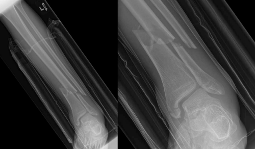
| A twenty-year-old male was admitted following an injury in a local public house, when a wall mounted radiator fell onto his left leg. Following initial assessment in the Accident and Emergency department, a fracture of the distal tibial metaphysis was identified. Radiographs revealed displaced fractures of the ipsilateral distal fibula and medial malleolus (Figure 1). He was transferred to theatre. |
| |
| |
|
|
Figure 1: Distal tibial metaphysis fracture and ipsilateral medial malleolus fracture. |
|
| |
| On a traction table, the fracture was reduced and stabilised with a T2 Stryker intramedullary nail. A standard medial para-patella approach was performed. An olive-tipped guidewire was inserted down the intramedullary canal of the tibia. Sequential reaming of the medullary canal facilitated the insertion of an appropriately sized intramedullary nail. Whilst proximal locking was performed using the jig, the distal locking was performed freehand under image intensifier guidance. |
| |
| A tension band fixation was fashioned to stabilise the medial malleolus fracture. The fracture site was exposed through a J-shaped incision, centred over the medial malleolus. The medial malleolus fracture was reduced and held with pointed reduction forceps. Two parallel 1.6 mm K-wires were inserted from the tip of the medial malleolus across the fracture site. A length of 1.2 mm wire was inserted beneath the deltoid ligament. The distal locking bolt of the intramedullary nail provided the proximal capstan of the tension band. A “Figure-of-8” tension band was adopted, with a double tension knot ( Figure 2). The 1.6 mm K-wires were then cut, bent and buried, to avoid irritation of the soft tissues. |
| |
|
|
Figure 2: Post-op AP and lateral radiographs showing intramedullary tibial nail and tension band wiring of the medial malleolus. |
|
| |
| Discussion |
| |
| We provide a novel approach to the management of a medial malleolus fracture in the presence of an ipsilateral fracture of the distal tibial metaphysis. |
| |
| Intramedullary tibial nailing is an established solution for managing fractures of the distal tibia and tension band wiring is an effective and widely used method for stabilising medial malleolus fractures, if the bony fragment is small or the bone is osteoporotic. |
| |
| To our knowledge, combining both techniques in this way to treat a distal metaphyseal tibial fracture and an ipsilateral medial malleolus fracture has not been previously described. |
| |
| The alternative to tension band wiring is the use of parallel, partially threaded cancellous screws. In this case, pre-operative planning had demonstrated that the position of the distal locking screw of the nail would interfere with the cancellous screws. The tension band technique reduces the risk of interference from the indwelling hardware. |
| |
| A different approach would be to use a MIPO technique. Reduction and stabilisation of the distal tibial fracture and medial malleolus can be addressed using a single LCP. |
| |
| One randomised controlled trial, comparing intramedullary nailing and MIPO for managing isolated distal tibial fractures concluded that nailing was the preferred option. The authors justified their conclusion by stating reduced operating time, reduced radiation exposure and ease of hardware removal, as their reasons for favouring intramedullary nailing. However, this statement is naturally contentious and open to debate by those who favour and have become more practised in MIPO. |
| |
| We believe that our technique provides a straightforward, easily reproducible and potentially cost effective answer to the problem of managing this injury pattern. |
| |
| |
| References |
| |
- Hamilton SW (2007) Parallel screw fixation of the medial malleolus. Ann R Coll Surg Engl 89: 729-730.
- AO Surgery Reference. www.aofoundation.org.
|
| |
| |


