| Research Article |
Open Access |
|
| Elena Marchetti1*, Andrea Guida2 and Stefano Eramo3 |
| 1School of Dentistry, University of Perugia, Lido di Ostia, Roma |
| 2Materials Science and Technology, University of Limerick, Roma |
| 3Conservative Dentistry Professor, University of Perugia, Roma |
| *Corresponding author: |
Elena Marchetti
University of Perugia
School of Dentistry
Via Sartena 900122
Lido di Ostia, Roma
Tel: 3281567485-065623973
E-mail: e.marchetti@libero.it |
|
| |
| Received December 15, 2011; Published September 28, 2012 |
| |
| Citation: Marchetti E, Guida A, Eramo S (2012) The Acquired Pellicle and the Enamel Etching: S.E.M. Finding. 1:338. doi:10.4172/scientificreports.338 |
| |
| Copyright: © 2012 Marchetti E, et al. This is an open-access article distributed under the terms of the Creative Commons Attribution License, which permits unrestricted use, distribution, and reproduction in any medium, provided the original author and source are credited. |
| |
| Abstract |
| |
| Aim: The aim of this study is to value the consequences made by the acquired pellicle on enamel etching. |
| |
| Methods and materials: Ten teeth without roots were sectioned in buccal-lingual direction at half crown; it has been obtained from each tooth two parts. Ten enamel specimens have been only cleansed on the buccal surface (group A) and ten enamel specimens (derived from the same teeth) have been polished with pumice powder and rotating brushes on its buccal surface (group B). All the specimens have been etched for 30 seconds and observed by SEM. The twenty images obtained were at first subjectively valued and compared in counterpart and randomized pairs by three independent operators which have expressed opinions following a scale. The microphotographies are also treated with Imaging Analysis (I.A.). |
| |
| Results: There are differences in the images of the etched enamel surfaces, which are from the same tooth, between buccal surface with acquired pellicle and the buccal surface without acquired pellicle. The I.A. showed a statistical difference (ANOVA test) in the extension of etched areas both between groups A and B and into. |
| |
| Conclusions: The acquired pellicle removal through a dental prophylaxis technique is necessary before the enamel etching since improve the effectiveness of this and, most probably, enable a better adhesion. |
| |
| Keywords |
| |
| Acquired pellicle; Etching; Enamel |
| |
| Introduction |
| |
| A good enamel cleaning before acid etching is necessary to prepare esthetic and functional direct and indirect restorations and fissure sealants [1,2]. Plaque and discolorations are removed by dental prophylaxis technique with pumice powder or paste and rotating brush or small rubber cup [3], there are other techniques like air-flow and bicarbonate jet polisher, which are as operative as the others and faster than the others, but they could damage tissues and pollute surfaces [4-6]. Although the good hygienic conditions of the patients (for example, patients who have done dental prophylaxis in the previous days) it is always necessary to remove the invisible acquired pellicle with dental prophylaxis. The acquired pellicle is a shapeless, organic and without cells pellicle, which covers just cleaned dental surfaces in a few minutes [7]. |
| |
| Acquired pellicle has a considerable importance for the production of tooth decay [8] especially for enamel demineralization/ remineralisation process [9,10] but the acquired pellicle could have an important place in the enamel surface response to no-bacterial acid exposition. |
| |
| There is a considerable literature about corrosive effects on enamel of acid drinks and substances, on one side acquired pellicle could be a barrier for the diffusion or a semi-permeable membrane between acids and dental surfaces [11,12] and on the other side it has been proved that acquired pellicle has an inhibition effect on enamel demineralization [13-15]. |
| |
| It has been rightly affirmed that “ in clinical conditions the effectiveness of acquired pellicle ability to protect dental surface is unknown, and the dental surface response to different acids exposure is unknown too” [16]. |
| |
| The acid orthophosphoric etching action to obtain the adhesion between enamel and resin seems to be the longest and the strongest on enamel surface. This in vitro study evaluates the difference on enamel etching between the same tooth surface with and without acquired pellicle. |
| |
| Materials and Methods |
| |
| Ten posterior teeth pull out for parodontal reasons and without caries, cleaned and sterilized in glutaraldehyde to avoid organic component damages have been used. The teeth without roots were sectioned in buccal-lingual direction at half crown; it has been obtained from each tooth two parts. For each tooth the buccal surface of one sample (group A) has been cleaned with toothpaste and brush, the buccal surface of the other sample (group B) of the same tooth has been cleaned with pumice powder and rotating brush. It has been obtained ten samples from group A, from A1 to A10, in which there was still acquired pellicle and ten samples from group B, from B1 to B10 (from the same teeth and with the same numbering of group A) which were without acquired pellicle. |
| |
| Samples of both groups were etched by 37% acid orthophosphoric gel for 30 seconds in a circular area with 2 mm diameter and rinsed with copious air-water jet. The obtained surfaces were prepared with a facing of one gold atomic layer and continually observed and photographed by SEM (Stereoscan Cambridge) at 5000X to observe possible differences in etching effectiveness. |
| |
| The twenty immages obtained were at first subjectively valued and compared in counterpart and randomized pairs by three independent operators which have expressed opinions following this scale: |
| |
| 4. Evident etching |
| |
| 3. Moderate etching |
| |
| 2. Scarce etching |
| |
| 1. Dubious etching |
| |
| 0. Void etching |
| |
| The obtained values were summarized in the table following the couples numbering. |
| |
| The same images were computed by a software for immages analysis (ImageJ, Scion Corp, USA) by which quantitative analysis and the comparison between the extensions of areas nocked by acid etching have been done. |
| |
| Softwer program (SPSS 11.0, SSPS Inc, USA) has been used for the statistical analysis, considering etching enamel surfaces as statistical units. Analysis of variance ANOVA has been used for the comparison between groups and into the group. |
| |
| Results |
| |
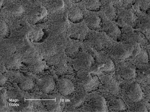
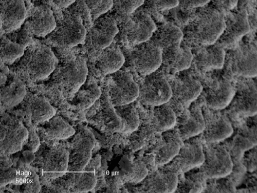
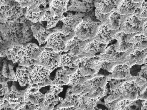
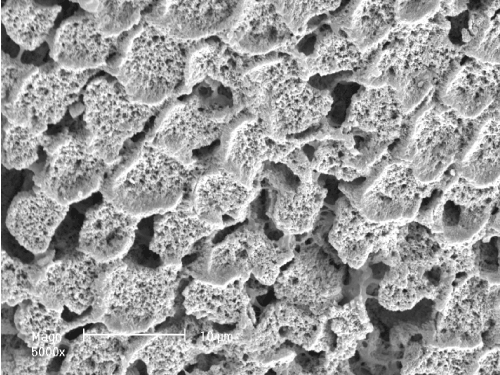
| The results are visible in twenty microelectronic immages, these are six examples: (SEM 5000X)-This is the buccal surface of A2 sample (with acquired pellicle), which has been etched. There's a moderate acid orthophosphoric etching around the enamel prisms and all the prisms' heads haven't been exposed (Figure 1). (SEM 5000X)- This is the buccal surface of B2 sample (without the acquired pellicle), which has been etched. This sample belongs to the same tooth of figure 1. There has been a better acid orthophosphoric etching than sample A2. In this sample there is an evident acid orthophosphoric etching around the enamel prisms and the prisms' heads have been well exposed (Figure 2). |
| |
|
|
Figure 1: (SEM, 5000X)-Enamel surface with acquired pellicle (sample A2), etched. |
|
| |
|
|
Figure 2: (SEM 5000X) - The same enamel surface without acquired pellicle (sample B2), etched. |
|
| |
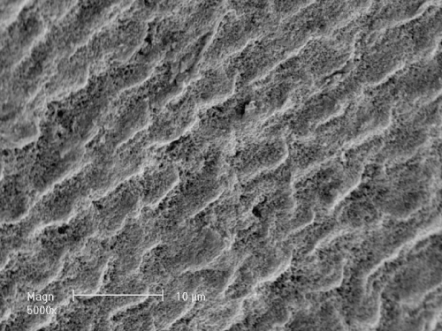
| (SEM 5000X)-This is the buccal surface of A6 sample (with acquired pellicle), which has been etched. There is a scarce acid orthophosphoric etching in enamel prisms and between prisms (Figure 3). |
| |
|
|
Figure 3: (SEM, 5000X)-Enamel surface with acquired pellicle (sample A6), etched. |
|
| |
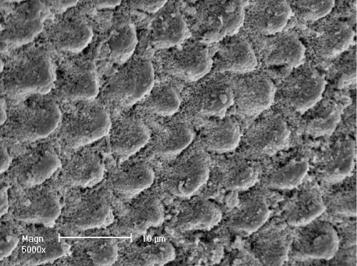
| (SEM 5000X)-This is the buccal surface of B6 sample (without the acquired pellicle) which has been etched. This sample belongs to the same tooth of figure 3. In this sample there's a moderate acid orthophosphoric etching in particular between the enamel prisms. There has been a better acid orthophosphoric etching than sample A6. The prisms' heads have been well exposed (Figure 4). |
| |
| |
|
|
Figure 4: (SEM 5000X)-The same enamel surface without acquired pellicle (sample B6), etched. |
|
| |
| (SEM 5000X)-This is the buccal surface of A9 sample (with the acquired pellicle), which has been etched. On this surface there is a demineralization area, which was evident before the acid etching too. In this case it isn't possible to value the acid etching effect on the sample (Figure 5). |
| |
|
|
Figure 5: (SEM, 5000X)-Eroded enamel surface with acquired pellicle (sample A9), etched. |
|
| |
| (SEM 5000X)-This is the buccal surface of B9 sample (without acquired pellicle), which has been etched. This sample belongs to the same tooth of figure 5. In this image the acid penetration is clearly more visible than in the previous image. The areas into the prisms have been etched in figure 5 as in figure 6, but the areas between prisms are larger and deeper than in this sample (Figure 6). |
| |
|
|
Figure 6: (SEM 5000X)-The same enamel surface without acquired pellicle (sample B9), etched. |
|
| |
| In table 1 there are the subjective valuations of the three independent operators: there are more etching evidences in all the B samples (without the acquired pellicle) than in A samples (also between 9A and 9B samples which are not clear). |
| |
|
|
Table 1: Opinions expressed by three independent operators on the sample images. |
|
| |
| Discussion |
| |
| There is only an experimental study on this argument besides ours and they obtained the same subjective results [17]. The Imaging Analysis on the twenty microelectronic 5000X images and their processing have proved that there is a statistical difference (ANOVA test) between the extensions of the enamel etched areas, in fact, enamel etched areas of B samples are bigger than the A samples (the B samples mean is 9.7% bigger than A samples) and between the samples of the same tooth, the B sample is bigger than A sample. |
| |
| Conclusion |
| |
| In patients which have a good oral hygiene condition (for example, if they have done a dental prophylaxis in the previous days) and need aesthetic fillings, the enamel cleaning could be neglected. |
| |
| The subjective valuations of these images and the objective results of imaging analysis have demonstrated that the acquired pellicle removal by dental prophylaxis technique is necessary before the enamel etching procedures. Acquired pellicle removal significantly improves (about 10%) the extension and depth of enamel etching, which is involved in the adhesive bonding according to literature. |
| |
| |
| References |
| |
- Cerutti A, Mangani G, Putignano A (2007) Odontoiatria estetica adesiva-Quintessenza Ed Int.
- Sol E, Espasa E, Boj JR, Canalda C (2000) Effect of different prophylaxis methods on sealant adhesion. J Clin Pediatr Dent 24: 211-214.
- Rios D, Honório HM, Francisconi LF, Magalhães AC, de Andrade Moreira Machado MA, et al. (2008) In situ effect of an erosive challenge on different restorative materials and on enamel adjacent to these materials. J Dent 36: 152-157.
- Boyde A (1984) Airpolishing effects on enamel, dentine, cement and bone. Br Dent J 156: 287-291.
- Negri PL, Eramo S e Lotito M (1991) Air flow e tessuti dentali. Doctor OS 8: 30-37.
- Honório HM, Rios D, Abdo RC, Machado MA (2006) Effect of different prophylaxis methods on sound and demineralized enamel. J Appl Oral Sci 14: 117-123.
- Dawes C, Jenkins GN, Tonge CH (1963) The nomenclature of the integuments of the enamel surface of the teeth. Br Dent J 115: 65-68.
- Tinanoff N (1976) The significance of the acquired pellicle in the practice of dentistry. ASDC J Dent Child 43: 20-24.
- Zahradnik RT, Moreno EC, Burke EJ (1976) Effect of salivary pellicle on enamel subsurface demineralization in vitro. J Dent Res 55: 664-670.
- Zahradnik RT (1979) Modification by salivary pellicles of in vitro enamel remineralization. J Dent Res 58: 2066-2073.
- Hannig M (1999) Ultrastructural investigation of pellicle morphogenesis at two different intraoral sites during a 24-h period. Clin Oral Investig 3: 88-95.
- Sonju Clasen AB, Hannig M, Sonju T (2000) Variations in pellicle thickness: a factor in tooth wear? In: Tooth wear and sensitivity:clinical advances in restorative dentistry. Addy M, Embery G , Edgar WM, Orchardson R (eds.). London: Taylor & Francis 189-195.
- Meurman JH, Frank RM (1991) Scanning electron microscopic study of the effect of salivary pellicle on enamel erosion. Caries Res 25: 1-6.
- Amaechi BT, Higham SM, Edgar WM, Milosevic A (1999) Thickness of acquired salivary pellicle as a determinant of the sites of dental erosion. J Dent Res 78: 1821-1828.
- Hannig M, Balz M (2001) Protective properties of salivary pellicles from two different intraoral sites on enamel erosion. Caries Res 35: 142-148.
- Hara AT , Ando M, González-Cabezas C, Cury JA, Serra MC, et al. (2006) Protective effect of the dental pellicle against erosive challenges in situ. J Dent Res 85: 612-616.
- Hoeppner MG, Sundfeld RH, Holland Jr C, Sundfeld MLMM (1998) Microscopic analysis of the in vitro penetration of a pit and fissure sealant on human dental enamel: effects of prophylaxis and times condizionamento enamel acid. Rev Bras Odontol 55: 258-264.
|
| |
| |

