| Research Article |
Open Access |
|
| J. Saraniya Devi1,2 and B. Valentin Bhimba2* |
| 1Department of Microbiology, Asan Memorial College of Arts and Science, Chennai-100, India |
| 2Department of Biotechnology, Sathyabama University, Chennai, India |
| *Corresponding authors: |
B. Valentin Bhimba
Department of Microbiology
Asan Memorial College of Arts and Science
Chennai-100, India
E-mail: bvbhimba@ yahoo.co.in |
|
| |
| Received February 03, 2012; Published August 24, 2012 |
| |
| Citation: Devi JS, Bhimba BV (2012) Anticancer Activity of Silver Nanoparticles Synthesized by the Seaweed Ulva lactuca Invitro. 1: 242. doi:10.4172/scientificreports.242 |
| |
| Copyright: © 2012 Devi JS, et al. This is an open-access article distributed under the terms of the Creative Commons Attribution License, which permits unrestricted use, distribution, and reproduction in any medium, provided the original author and source are credited. |
| |
| Abstract |
| |
| Introduction: A novel process for the rapid synthesis of AgNPs using the aqueous extract of marine macroalgae, Ulva lactuca. |
| |
| Methods: The nanoparticles were synthesized by subjecting the reaction media at 121°C for 10 min. and were assessed for in vitro cytotoxic activity on Hep-2, MF7, HT29 and Vero cell lines. |
| |
| Results: The synthesized AgNPs were characterized by observing a peak at 434nm using UV-visible spectroscopy. Fourier Transform Infra Red spectroscopic analysis was carried out to identify the functional groups responsible for the synthesis of AgNPs. The synthesized AgNPs were further demonstrated and confirmed by the characteristic peaks observed in the XRD image. SEM and TEM images revealed that AgNPs were spherical in shape and measured 20-56nm in size. Qualitative as well as quantitative status of elements that might be involved in the formation of AgNPs is stressed by EDAX analysis. |
| |
| Conclusion: The synthesized nanoparticles were potently cytotoxic against Hep 2 cell lines and mildly cytotoxic against MCF 7 and HT 29 cell lines. The synthesized AgNPs were found to be a cytotoxic against human (Hep2, MCF7 and HT29) cancer cell lines. |
| |
| Keywords |
| |
| Silver nanoparticles; Marine macroalgae-Ulva lactuca; In vitro cytotoxicity; Hep2 cell lines; MCF7 cell lines; HT29 cell lines; Vero cell lines |
| |
| Introduction |
| |
| With the terrestrial resources being greatly explored and exploited, researchers turn to the oceans for numerous reasons. The oceans cover more than 70% of the world surface housing 34 living phyla out of the 36 and more than 300,000 known species of fauna and flora. The marine environment is known to contain over 80% of world’s plant and animal species. In recent years, many bioactive compounds have been extracted from various marine plants, marine animals and marine organisms [1]. |
| |
| Seaweeds are marine plants because they use the sun's energy to produce carbohydrates from carbon dioxide and water. In India seaweeds are mainly exploited by industries for phycocolloids but very poorly explored for their beneficial application in pharmacology [2]. Seaweeds are important sources of protein, iodine, vitamins, minerals [3] and polyphenols [4] with their metabolites showing wide range of promising biological activities. Over the past several decades, seaweeds and their extracts have generated an enormous amount of interest in the pharmaceutical industry as a fresh source of bioactive compounds with immense medicinal potential [5]. The macro algae reported to contain various significant compounds with antibacterial [6], antiviral [7] and antitumor activity [8]. AgNPs have been synthesized using marine microalgae and screened for its antibacterial activity [9]. |
| |
| Discovery of anticancer drugs that must kill or disable only tumor cells without undue toxicity is an extraordinary challenge [10]. Toxicity of plant or microbial material is considered as the presence of antitumor compounds [11]. In recent years, an increasing number of marine natural products have been reported to display antimicrobial activities [12] and anti-tumor compounds have been isolated from sponges, tunicates, algae and other organisms [13]. In most cases, the evaluation of the anti-cancer potential of crude extracts from different sea organisms has been carried out by in vitro cytotoxicity tests in malignant cell cultures [14]. Hundreds of potential anti-tumor agents have been isolated from marine origin especially from marine algae [15,16]. Isolation of cytotoxic anti-tumor substances from marine organisms has been reported in several references during the last 40 years [17]. |
| |
| In this study, AgNPs were synthesized using Ulva lactuca and its cytotoxicity was assessed against Hep-2, HT-29, MCF-7 and Vero cell lines. |
| |
| Methods |
| |
| Seaweed |
| |
| Fresh seaweed sample were collected in polythene bags from the intertidal regions of the Mandapam coastal regions (Lat.09ÃÅ¡ 17.417 N; Long.079ÃÅ¡ 08.558 E) of Gulf of Mannar. Samples were cleaned thoroughly by washing with running tap water to remove stones, epiphytes, extraneous matter and necrotic parts. Then cut into small pieces and rinsed with sterile distilled water. The cleaned macro-algae were shade dried at room temperature for a week. The shade-dried thalli were powdered and aqueous extract was prepared. |
| |
| Rapid synthesis of AgNPs |
| |
| 1gm of seaweed powder was extracted with water and filtered. 1mM silver nitrate solution was added to the filtrate slowly under magnetic stirring conditions for even coating of silver and subjected to heating at 12°C for 10 min. The extract is used as reducing and stabilizing agent for 1mM of Silver nitrate. This one pot green synthesis was the modified method followed by Vigneshwaran et al. [18]. |
| |
| Characterization of AgNPs |
| |
| Through sampling the bioreduction of Ag+ in aqueous solution of seaweed extract was monitored. Diluted 0.2 ml reaction media with 2 ml deionized water and the resulting diluents were subsequently measured by UV-Vis spectra. UV-Vis spectroscopy analyses of silver nanoparticles produced were carried out as a function of bioreduction time on Ultraviolet-Visible spectroscopy (UV-1601 pc shimadzu spectrophotometer). |
| |
| Samples of the aqueous solution of the silver nanoparticles (AgNPs) were prepared by centrifugation at 10,000 rpm for 30 min. The pellet was lyophilized and subjected to FT-IR analysis by KBR pellet (FT-IR grade) method and the spectrum was recorded using spectral range of 4000~400 cm-1 (Thermo Nicolet, Avatar 370). |
| |
| The interaction between protein-silver nanoparticles were analyzed by Fourier transform infrared spectroscopy (FTIR) in the diffuse refluctance mode at a resolution of 4cm-1 in the KBr pellets and the spectra were recorded in the wavelength interval of 4000 to 400nm-1. FTIR measurements were carried out to identify the biomolecules responsible for the reduction of the Ag+ ions and the capping of the bioreduced AgNPs synthesized by seaweed extract. For comparative study, the seaweed filtrate was mixed with KBr powder, pelletized after drying and subjected to measurement. |
| |
| X-ray diffraction (XRD) measurement of the seaweed reduced AgNPs was carried out using powder X-ray diffractometer instrument (PXRD-6000 SCHIMADZU) in the angle range of 10°C-80°C at 2θ, scan axis: 2:1 sym. The size of the AgNPs was calculated from the PXRD peak positions using Bragg’s law. |
| |
| Thin films of the sample were prepared on a carbon coated copper grid by dropping a minute of the sample on the grid. The film on the SEM grid were allowed to dry under a mercury lamp for 5 min prior to measurement using Quanta 200 FEG SEM machine. |
| |
| The presence of elemental Silver was confirmed through EDAX. The EDAX observations were carried out in STIC, CUSAT, Kerala (JOEL Model JED-2300). The EDAX spectrum recorded in the spotprofile mode from one of the densely populated silver nanoparticles region on the surface of the film. The nanocrystallites were analyzed using Quanta 200 FEG. |
| |
| Sample for Transmission Electron Microscopic (TEM) analysis were prepared by coating AgNPs solutions onto carbon coated copper TEM grids. The film on the TEM grid was allowed to dry prior to measurement using TECHNAI 10 instrument operated at an accelerating voltage at 80KV. |
| |
| Evaluation of in vitro cytotoxic activity of the silver nanoparticles on cell lines |
| MTT assay was performed to determine the cytotoxic property of synthesized AgNPs against Hep-2, MF7, HT29 and Vero cell lines. Cell lines were seeded in 96-well tissue culture plates. Stock solutions of AgNPs (5 mg/ml) were prepared in sterile distilled water and diluted to the required concentrations (10, 5, 2.5, 1.25, 0. 625, 0.312, 0.156 mg/ml) using the cell culture medium. Appropriate concentrations of Ag-NP stock solution were added to the cultures to obtain respective concentration of Ag-NP and incubated for 48 hrs at 37°C. Non-treated cells were used as control. Incubated cultured cell was then subjected to MTT (3-(4,5-Dimethylthiazol-2-yl)-2,5-diphenyltetrazolium bromide, a tetrazole) colorimetric assay [19]. The tetrazolium salt 3-[4,5-dimethylthiazol-2-yl]-2,5-diphenyltetrazolium bromide (MTT) is used to determine cell viability in assays of cell proliferation and cytotoxicity. MTT is reduced in metabolically active cells to yield an insoluble purple formazon product. Cells were harvested from maintenance cultures in the exponential phase and counted by a hemocytometer using trypan blue solution. The cell suspensions were dispensed (100μl) in triplicate into 96-well culture plates at optimized concentrations of 1 × 10⁵/well for each cell lines, after a 24- hr recovery period. Assay plates were read using a spectrophotometer at 520 nm. The spectrophotometrical absorbance of the samples was measured using a microplate (ELISA) reader. The cytotoxicity data was standardized by determining absorbance and calculating the correspondent AgNP concentrations.Data generated were used to plot a dose-response curve of which the concentration of extract required to kill 50% of cell population (IC50) was determined. |
| |
| Cell viability (%) = Mean OD/ control OD × 100 |
| |
| Following Ag-NP treatment, the plates were observed under an inverted microscope to detect morphological changes and photographed. |
| |
| Results and Discussion |
| |
| Marine algae (seaweeds) contain a number of biodynamic compounds of therapeutic value. These compounds provide valuable ideas for the development of new drugs against cancer, microbial infections and inflammations [20]. Many of the secondary metabolites biosynthesized by the marine plants are well known for their cytotoxic property [21]. |
| |
| The present study was conducted to screen the marine macro-algae, Ulva lactuca for the synthesis of AgNPs and its cytotoxic potential against cancer cell lines. Reduction of silver ions into AgNPs during exposure of the seaweed extracts could be followed by color change when kept in an autoclave at 121°C for 10min. The reaction mixture turning brown after the addition of silver nitrate solution is a clear indication of the formation of silver nanoparticles [22]. Simultaneously control without silver ions was also run along with the experimental flask [23]. |
| |
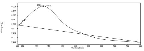
| UV vis absorption spectra are known to be quite sensitive to the formation of AgNPs. Thus the presence of AgNPs was characterized by using a UV-Vis spectrum [24]. A single broad peak was observed at 434 nm (Figure 1), which corresponds to plasmon excitation of the AgNPs. A control without the addition of the silver nitrate solution was also recorded. Several investigators have observed absorption maxima of colloidal silver solution between 410 to 440 nm, which is assigned to surface plasmon of various metal nanoparticles [25]. |
| |
| |
|
|
Figure 1: UV visible spectra of synthesized AgNPs against the aqueous extract of Ulva latuca. |
|
| |
| FTIR measurements were carried out to identify biomoloecules responsible for the stabilization of the newly synthesized AgNPs. The FT-IR spectrum of the seaweed extract in Figure 2a showed the absence of AgNO3 with transmission peaks at 3431.08, 2172.25 & 1747.83 cm-1 and Figure 2b shows the presence of AgNO3 with transmission peaks at 3661.86, 3539.98, 2083.96 & 1659.46 cm-1 respectively. The band at 3661.86 is corresponded to alcohol, phenol O-H, free group. The band at 3539.98 corresponded to an amide N-H band; band at 2083.96 corresponded to alkyne C-C and the band 1659.46 corresponded to amide C=O group. This evidence suggested the release of protein molecules that probably had a role in the formation and stabilization of AgNPs in aqueous solution. |
| |
|
|
Figure 2: a) FT-IR spectrum of aqueous extract of b) FT-IR spectrum of synthesized AgNPs Ulva latuca. |
|
| |
| The synthesized AgNPs was further demonstrated and confirmed by the characteristic peaks observed in the XRD image (Figure 3). The sharp diffraction patterns of the XRD spectra obtained by the annealing at 200°C indicates a pure crystalline silver structure, JCPDS card no: 04-0783. The figure shows 3 peaks at 2θ values of 37.77, 44.15 and 65.25 corresponding to 111, 200 and 220 planes of silver respectively. We observed no impurity peak in the X-ray diffraction pattern. All diffraction peaks correspond to the characteristic face centered cubic (FCC) phase [26]. The X-rays are scattered by diffraction owing to the unique crystalline structure of the material analyzed. From this, crystalline structure of the material can be obtained. Sambhy et al used this analytical method to characterize the particle structure and confirm the presence of the nanoparticles [27]. |
| |
|
|
Figure 3: XRD pattern of synthesized AgNPs. |
|
| |
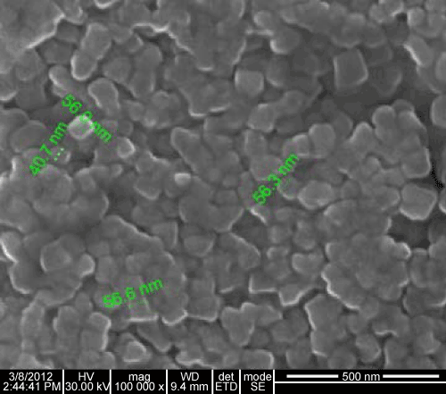
| The SEM micrographs of AgNPs obtained showed that they are spherical shaped, well distributed in solution with an average size of about 56nm in Figure 4. Similar phenomenon was reported by Chandran et al. [19]. |
| |
|
|
Figure 4: SEM images of synthesized AgNPs. |
|
| |
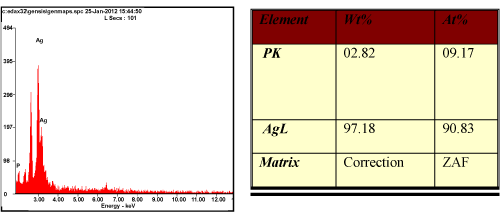
| Energy Dispersive Analysis of X-ray (EDAX) gives qualitative as well as quantitative status of elements that may be involved in the formation of AgNPs. Figure 5 shows the peak in silver region at 3KeV which is typical for the absorption of metallic silver nanocrystalline due to surface plasmon resonance. The presence of strong signals from silver (90.83%) atoms in the nanoparticles and weaker signals from phosphorous (9.17%) atoms was thus confirmed. The P, O signals are likely to be due to X ray emission from proteins/enzymes present in the seaweed [28]. |
| |
|
|
Figure 5: EDAX profiles of AgNPs along with the elemental composition of Ag. |
|
| |
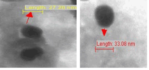
| A TEM micrograph recorded from the silver nanoparticles deposited on carbon coated copper TEM grid was shown in Figure 6. This micrograph shows spherical AgNPs with low density dispersion and are in the range of 20-56nm in size. Characterization of nanopartiles by TEM has been reported by Sondi and Salopek-Sondi [29]. |
| |
|
|
Figure 6: TEM images of synthesized AgNPs. |
|
| |
| In vitro cytotoxic activity against Hep2, MCF7, HT29 and Vero cell line at different concentrations was evaluated and compared with the standard drug 5-fluorouracil. The in vitro screening of the AgNPs showed potential cytotoxic activity against the human laryngeal cancer (Hep-2) cell line, human breast cancer (MCF 7) cell line and human colon cancer (HT 29) cell line. Less cytotoxicity of synthesized AgNPs against normal Vero cell line with IC50=95μg/ml are shown in Table |
| |
|
|
Table 1: Results of the MTT assay test on Hep 2, MCF 7, HT 29 and Vero cell lines after treatment with conce ntrations of synthesized AgNPs. |
|
| |
| 1. The plates were observed under an inverted microscope to detect morphological changes. The result showed that Hep2 cells proliferation were significantly inhibited by AgNPs with an IC50 value of 12.5μg/ml of the concentration, MCF7 cells with an IC50 value of 37μg/ml of the concentration and HT29 cells with an IC50 value of 49μg/ml of the concentration. Thus the synthesized nanoparticles were found to be potently cytotoxic agent against Hep 2 cell lines and mildly cytotoxic against MCF 7 and HT 29 cell lines. These results indicate that the sensitivity of human cancer cell line for cytotoxic drugs is higher than that of Vero cell line for the same cytotoxic agents. |
| |
| There are reports that marine macroalgae belonging to Phaeophyta group possess antitumor activity, and sterols from Sargassum carpophyllum exhibited cytotoxic activity against several cultured cell lines [30]. Several cytotoxic compounds such as fucoidans, laminarians, and terpenoids stated to posses anticancer, antitumor, antibacterial and anti-proliferative properties are reported to be abundant in seaweeds [31]. These compounds could be further explored as novel leads to cancer chemoprevention and chemotherapy and necessitates further investigation [32]. In present cancer claims the lives of approximately seven million people worldwide on annual basis. Hence, in recent years, the search for the cancer therapeutics from natural products has been on. Bioactive compounds in marine plants have been reported against various cancer cell lines. Cytotoxic effect is inversely proportional to the size of the bioactive compound AgNP. |
| |
| Conclusion |
| |
| Silver nanoparticles were synthesized by a rapid synthetic method from a marine macro alga, Ulva lactuca. The synthesized nanoparticles were characterized and were tested for its efficiency as a potent cytotoxic agent against human cancer cell lines. In this study, it was observed that the synthesized AgNPs induces a concentration dependent inhibition of cells. Almost 60% of drugs approved for cancer treatment are of natural origin and there is an urgent need and hope to develop alternative therapeutic measures against this deadly disease. |
| |
| |
| References |
| |
- Bhimba VB, Meenupriya J, Joel EL, Naveena ED, Kumar S, et al. (2010) Antibacterial activity and characterization of secondary metabolites isolated from mangrove plantAvicennia officinalis. Asian Pac J Trop Med 3: 544-546.
- Kaliaperumal N, Kalimuthu S, Ramalingam JR (2004) Present scenario of seaweed exploitation and industry in India. Seaweed Res Utiln 26: 47-53.
- Mans DR, da Rocha AB, Schwartzman G (2000) Anti-cancer drug discovery and development in Brazil, targeted plant collection as a rational strategy to acquire candidate anticancer compounds. Oncologist 5: 185-198.
- Yoshie Y, Wang W, Hsieh YP, Suzuki T (2002) Compositional difference of phenolic compounds between two seaweeds, Halimeda spp. J Tokyo Univ Fish 88: 21-24.
- Smith AJ (2004) Medical and pharmaceutical applications of seaweed natural products-a review. J Appl Phycol 16: 245-262.
- Nair R, Chabhadiya R, Chanda S (2007) Marine algae: Screening for a potent antibacterial agent. J Herb Pharmacother 7: 73-86.
- Richards JT, Kern ER, Glasgow LA, Overall JC, Deign EF, et al. (1978) Antiviral activity of extracts from marine algae. Antimicrobial Agents Chemother 14: 24-30.
- Espeche ME, Fraile ER, Mayer AMS (1984) Screening of Argentine marine algae for antimicrobial activity. Hydrobiology 116: 525- 528.
- Merin DS, Prakash, Bhimba VB (2010) Antibacterial screening of silver nanoparticles synthesized by marine microalgae. Asian Pac J Trop Med 3: 797-798.
- Hameed SV, Sultana J, Ara S, Ehteshamul-Haque, Athar M (2009) Toxicity of Fusarium solanistrains on Brine shrimp (Artemia salina). Zoological Research 30: 468-472.
- Bhimba VB, Franco DA, Mathew JM, Jose GM, Joel EL, et al. (2012) Anticancer and antimicrobial activity of mangrove derived fungi Hypocrea lixii VB1. Chin J Nat Med 10: 77-80.
- Joel EL, Bhimba VB (2010) Isolation and characterization of secondary metabolites from the mangrove plant Rhizophora mucronata. Asian Pac J Trop Med 3: 602-604.
- Chapman DJ, Gellenbeck KW (1983) An historical prespective of algal biotechnology. In: “Algae and cyanobacterial biotechnology”, Longman group, UK.
- Russell FE (1963) Advances of marine biology. Academic press, New York.
- Adams NM (1994) Seaweeds of New Zealand: An Illustrated Guide. Canterbury University Press, Christchurch, New Zealand.
- Fadli M, Aracil JM, Jeanty G, Banaigs B, Francisco C (1991) Novel Meroterpnoids from Cystoseria mediterranea: use of the crown-gall bioassay as a primary screen for lipophilic anti-neoplatic agents. J Nat Prod 54: 261-264.
- Gonzalez AG, Darias V, Estevez E (1997) Chemo-Therapeutic activity of polyhalogenated terpenes from Spanish algae. Planta Med 44: 44-46.
- Vigneshwaran N, Nachane RP, Balasubramanya RH, Varadarajan PV (2006) A novel one-pot ‘green’ synthesis of stable silver nanoparticles using soluble starch. Carbohydr Res 134: 2012-2018.
- Chandran SP, Chaudhary M, Pasricha R, Ahmad A, Sastry M (2006) Synthesis of gold nanotriangles and silver nanoparticles using Aloe vera plant extract. Biotechnol Prog 22: 577-583.
- Selvin J, Lipton AP (2004) Biopotentials of Ulva fasciata and Hypnea musciformiscollected from the peninsular coast of India. J Mar Sci Tech 12: 1-6.
- Manilal A, Sujith S, Kiran GS, Selvin J, Shakir C, et al. (2009) Bio-potentials of seaweeds collected from Southwest coast of India. J Mar Sci & Technol 17: 67-73.
- Ahmad A, Senapati S, Khan IM, Kumar R, Ramani R, et al. (2003) Intracellular synthesis of gold nanoparticles by a novel alkalotolerant actinomycete, Rhodococcus species. Nanotechnology 14: 824-828.
- Ray S, Sarkar S, Kundu S (2011) Extracellular Biosynthesis of Silver Nanoparticles Using The Mycorrhizal Mushroom Tricholoma Crassum (Berk.) Sacc.: Its Antimicrobial Activity Against Pathogenic Bacteria And Fungus, Including Multidrug Resistant Plant And Human Bacteria. Digest Journal of Nanomaterials and Biostructures 6: 1289-1299.
- Jain P, Pardeep T (2005) Potential of silver nanoparticle-coated polyurethane foam as an antibacterial water filter. Biotechnol Bioeng 90: 59-63.
- Sharma VK, Yngard RA, Lin Y (2009) Silver nanoparticles: Green synthesis and their antimicrobial activities. Advances in Colloids and Interface Science 145: 83-96.
- Leff DV, Brandt L, Heath JR (1996) Synthesis and characterization of Hydrophobic, organically soluble gold nanocrystals functionalized with primary amines. Langmuir 12: 4723-4730.
- Sambhy V, MacBride MM, Peterson BR, Sen A (2006) Silver bromide nanoparticle/polymer composites: Dual action tunable antimicrobial materials. J Am Chem Soc 128: 9798-9808.
- Xu H, Kall M (2002) Modeling the optical response of nanoparticle-based surface plasmon resonance sensors. Sensors and Actuators B: Chemical 4: 244-249.
- Sondi I, Salopek-Sondi B (2004) Silver nanoparticles as antimicrobial agent: a case study on E. coli as a model for Gram-negative bacteria. J Colloids Interface Sci275: 177-182.
- Tang HF, Yang-Hua Y, Yao XS, Xu QZ, Zhang XY, et al. (2002) Bioactive steroids from the brown alga Sargassum carpophyllum. J Asian Nat Prod Res 4: 95-105.
- Smit AJ (2004) Medicinal and pharmaceutical uses of seaweed natural products: A review. J Appl Phycol 16: 245-262.
- Vinayak RC, Sabu AS, Chatterji A (2011) Bioprospecting of a few brown seaweed for their cytotoxic and antioxidant activities. Evidence-Based Complementary and Alternative Medicine: 1-9.
|
| |
| |


