| Research Article |
Open Access |
|
| Chandramani B More* and Hetul J. Patel |
| Department of Oral Medicine & Radiology,K. M. Shah Dental College & Hospital, Sumandeep Vidyapeeth, Piparia, Vadodara , Gujarat, India |
| *Corresponding authors: |
Chandramani B. More
Department of Oral Medicine & Hospital
Sumandeep Vidyapeeth, Piparia
Vadodara, Gujarat, India
Tel: +91 9974900278
E-mail: drchandramanimore@rediffmail.com |
|
| |
| Received April 02, 2012; Published July 23, 2012 |
| |
| Citation: More CB, Patel HJ (2012) Dens Invaginatus: A Radiographic Analysis. 1: 147. doi:10.4172/scientificreports.147 |
| |
| Copyright: © 2012 More CB, et al . This is an open-access article distributed under the terms of the Creative Commons Attribution License, which permits unrestricted use, distribution, and reproduction in any medium, provided the original author and source are credited. |
| |
| Abstract |
| |
| Background: Dens invaginatus is a malformation of teeth probably resulting from an infolding of the dental papilla during tooth development. The maxillary lateral incisor is the most commonly affected tooth. The malformation shows a broad spectrum of morphologic variations and frequently results in early pulp necrosis. The radiographic examination is the most realistic way to diagnose the invagination. |
| |
| Objective: To analyze clinical and radiographic records of Dens invaginatus with emphasis on radiographic findings. The present study describes the clinical and radiographic features related to the different types of dens invaginatus and highlights those features which may indicate the presence of a previously undetected invagination. |
| |
| Study design: The present hospital based retrospective study was conducted by assessing the clinical and radiographic records of year 2009 & 2010, available in the archives of the department. Seventy five dental radiographs formed the basis of this study. The data was evaluated and analyzed using Microsoft excel 2007. |
| |
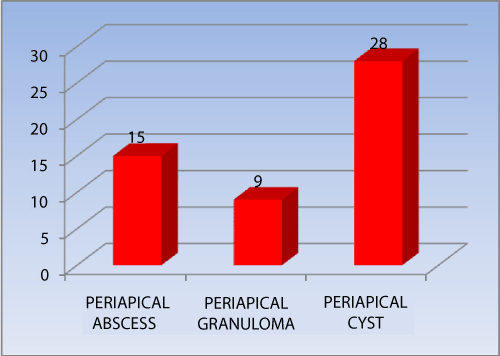
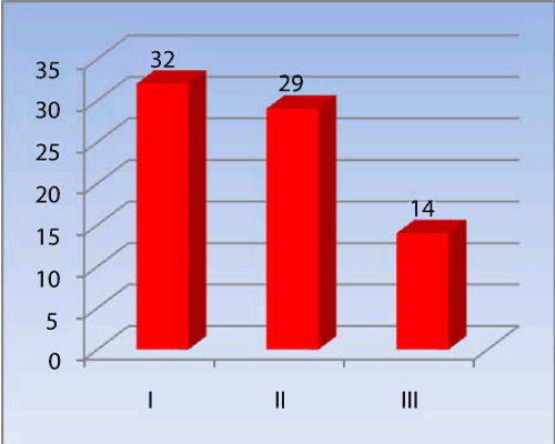
| Results: From the present study, it was concluded that dens invaginatus was observed in the age group of 16-40 years, seen in males and females with ratio of 1:1.2, more prevalent in maxillary lateral incisor (54.7%) and periapical lesions were noted in 69.3% [ abscess - 15 cases (20%), granuloma - 9 cases (12%) or cyst - 28 cases (37.33%) ]. It was distinctly noticed that maximum number of teeth were affected by type-I (42.7%), type- II (38.7%) & type- III (18.6%) invagination. |
| |
| Conclusion: Dens invaginatus is clinically significant due to the possibility of pulpal involvement. An early diagnosis of invagination is crucial and requires thorough clinical & radiographic examination of all teeth especially lateral incisors. |
| |
| Keywords |
| |
| Anomalous teeth; Dens invaginatus; Dens in dente; Radiographic analysis; Telescopictooth |
| |
| Introduction |
| |
| Dens Invaginatus (DI) is a dental anomaly which results from invagination of enamel organ into dental papilla, beginning at the crown and sometimes extending into the root before calcification [1]. This condition is also known as “Dens in Dente”, “Dilated composite odontome” “Gestant odontoma”, “Dentinoid in dente” or “Telescopic tooth” [2] Hallet introduced the term dens invaginatus in order to clarify the point that enamel is located centrally and the dentine peripherally due to the invagination. Since then it has been a preferred term, although dens in dente is a more commonly used term [1,2] . |
| |
| The involved tooth crown as well as root may exhibit variations in size and form which was first noticed in whale’s tooth by Ploquet in 1794.[3] Salter first described anatomical anomaly as “a tooth within tooth” in 1855 [4] and was the first person who described dens invaginatus in human tooth in 1856 [3]. The etiology is controversial and remains unclear. The possible factors responsible are, lateral fusion of two germs, constriction of dental arch in the enamel organ, increased external pressure, focal growth retardation, focal growth stimulation in certain areas of tooth buds, invagination of the crown before calcification of the teeth, infection and genetic factors [5-8]. |
| |
| Clinically, a morphologic alteration of the crown or a deep foramen coecum can serve as an indication for the diagnosis of dens invaginatus [5]. But affected tooth may not show clinical signs of the malformation. As maxillary lateral incisors are the teeth most susceptible to coronal invaginations, these teeth should be investigated thoroughly clinically and radiographically, atleast in all cases with a deep pit at the foramen coecum. If one tooth is affected in a patient, the contra-lateral tooth should also be investigated. The main reason for consultation is acute pain and inflammation. Most cases of dens invaginatus are detected after a routine radiographic evaluation with a panoramic x-ray and confirmed with a periapical film [6]. Thorough clinical and radiographic examination is required to diagnose and successfully treat minor to severe invaginations. Modern clinical techniques may facilitate the management of invaginations. |
| |
| The purpose of the present study was to analyse cases of dens invaginatus with emphasis on radiographic findings and to review the current literature on this dental anomaly briefly and discuss the clinical and radiographic findings that may aid in diagnosis and strategies for preventing loss of vitality. |
| |
| Methodology |
| |
| The present hospital based retrospective study was conducted by assessing the clinical and radiographic records of dens invaginatus between the year 2009 & 2010, available in the archives of department. The permission to undertake this study was obtained from the Institutional Ethics Committee. The total of seventy five teeth having dens invaginatus were part of the study. Out which sixty seven teeth were subjected to prophylactic invagination treatment. The intra oral periapical radiographs formed the basis of the present study. The radiographs with any type of artifact or fault were excluded from the study. All the radiographs were taken by using bisecting angle technique, were developed under standardized conditions and viewed on a standard illuminated screen by three oral radiologists to prevent inter observer bias. To determine the type of invagination, Ohlers’ system of classification for Dens invaginatus was used. The descriptive Data analysis was performed using Microsoft Office Excel 2007. The study variables included age, gender, affected tooth, any associated lesion and type of invagination. |
| |
| Results |
| |
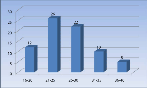
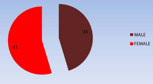
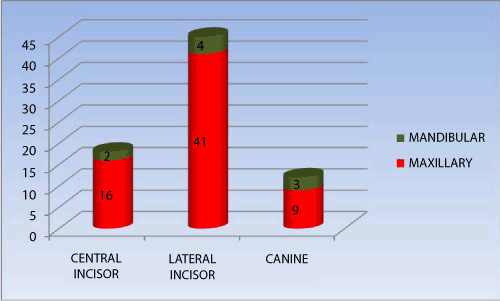
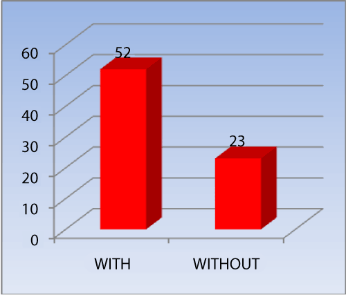
| In the present study we observed that, the patient’s affected with dens invaginatus belonged to the age group of 16-40 years with the mean age of 28 years (Graph 1). The male to female ratio was 1:1.2 (Graph 2). All the affected teeth were from permanent dentition. Out of total seventy five teeth, maxillary teeth (88%) were most affected as compared to the mandibular teeth (12%). It was also observed that, the most common tooth affected by dens invaginatus is maxillary lateral incisors (54.7%), followed by maxillary central incisor (21.33%), maxillary canine (12%), mandibular lateral incisors (5.33%), mandibular canine (4%) and mandibular central incisor (2.67%) (Graph 3). It was also noted that periapical lesions were present in 52 cases (69.3%) & absent in 23 cases (30.7%) (Graph 4). The periapical lesions were abscess (20%), granuloma (12%) or cyst (37.33%) (Graph 5). According to Ohler’s system of classification, the type- I pattern was seen in 32 cases (42.7%), type- II in 29 cases (38.7%) & type- III in 14 cases (18.6%) (Graph 6). |
| |
|
|
Graph 1: showing Age wise distribution. |
|
| |
|
|
Graph 2: Showing Gender wise distribution. |
|
| |
|
|
Graph 3: Showing tooth involved. |
|
| |
|
|
Graph 4: Showing involvement of periapical lesions. |
|
| |
|
|
Graph 5: Showing type of Periapical lesion. |
|
| |
|
|
Graph 6: Showing types of Dens Invagination. |
|
| |
| Discussion |
| |
| Dens Invaginatus (DI) occur rarely in primary but frequently in permanent dentition and has a general prevalence of 0.04–10%. In our study all the seventy five teeth belonged to permanent dentition. Literature study shows that, DI is commonly seen in maxillary teeth and involvement in mandibular teeth is rare [9]. This observation is consistent with the findings of the present study. We noted that 88% of the cases had maxillary teeth involvement. |
| |
| Maxillary lateral incisors are the teeth most susceptible to coronal invaginations [6]. Central incisors, premolars, canines and molars are in decreasing order of frequency. In our study it was observed that, the most common tooth affected by dens invaginatus is maxillary lateral incisors (54.7%), followed by maxillary central incisor (21.33%), maxillary canine (12%), mandibular lateral incisors (5.33%), mandibular canine (4%) and mandibular central incisor (2.67%). The reason behind this is thought to be the unfavorable position of maxillary lateral incisor during its formative stages and it is the last anterior tooth to calcify [10]. |
| |
| Bilateral occurrence of DI is not uncommon and occurs in 43% of all cases with a high degree of inheritance [3]. But our study did not show any bilateral occurrence of DI, may be due to small sample size. |
| |
| The gender predilection for DI i.e. male to female ratio is 1:1.7, with a slight female predilection [11]. Our study co-related this finding, wherein we observed that the male to female ratio was 1:1.2, with female predilection. |
| |
| Dens invaginatus is clinically significant due to the possibility of the pulp being affected. As pulpal involvement of teeth with coronal invaginations may occur shortly after tooth eruption, an early diagnosis is mandatory to instigate preventive treatment. Clinical examination may reveal a deep fissure or pit on the surface of an anterior tooth. Due to the tortuous lingual anatomy, it is possible for caries to develop inside the invagination without any clinically detectable lesion. Since the enamel lining is thin and in close proximity to the pulp chamber, a carious lesion could easily perforate the pulp chamber. Further, there are sometimes thin canals within the enamel of the dens invaginatus, forming a direct communication with the pulp. Hence pulpitis and necrotic pulps are often associated with this anomaly. The other reported sequelae of undiagnosed and untreated coronal invaginations are retention of neighboring teeth, displacement of teeth, cysts and internal resorption. In the present study we observed that, out of seventy five cases, 52 cases (69.33%) had periapical lesions either abscesses - 15 cases (20%), granuloma - 9 cases (12%) or cyst - 28 cases (37.33%) |
| |
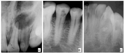
| Dens invaginatus is often a surreptitious finding. Upon radiographic evidence of a dens invaginatus, the apical periodontium should be examined. If the radiographic appearance is unremarkable, pulp sensitivity testing should be performed. If the results suggest vital and unaffected pulpal tissue, then the tooth should be promptly restored to curtail access of the dens invaginatus to the oral environment [5] (Figure 1). |
| |
|
|
Figure 1: A showing Type I Dens Invaginatus in lateral incisors. Note- periapical cyst. B Type II dens invaginatus in second premolar and C Type III dens invaginatus in mandibular canine. |
|
| |
| The Ohlers’s system is the most popularly used classification for DI. According to this system, invaginations are classified into the following three types based on the depth of the invagination and the degree of communication with the periodontal ligament or the peri-radicular tissue [3,12] |
| |
| Type I: The invagination ends in a blind sac, limited to the dental crown. |
| |
| Type II: The invagination extends to the cemento–enamel junction , also ending in a blind sac. |
| |
| Type III: The invagination extends to the interior of the root, providing an opening to the periodontium, sometimes presenting another foramen in the apical region of the tooth [13]. |
| |
| In this study, we distinctly observed that type I pattern was seen in 32 cases (42.7%), type II in 29 cases (38.7%) & type III in 14 cases (18.6%). |
| |
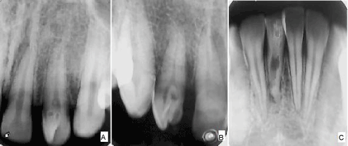
| Dens invaginatus are known to be associated with other abnormalities such as taurodontism, microdontia, talon cusp (Figure 2), gemination, supernumerary tooth, dentinogenesis imperfecta, short roots and with some medical–dental syndromes[12,14,15]. H. M. Worth has described two types of dens in dente: first is a root dilation resembling an open umbrella and other one is fleur-de-lys which resembles the French emblem [16]. The treatment of dens invaginatus ranges from preventive and restorative treatment procedures to nonsurgical root canal or surgical treatment. |
| |
|
|
Figure 2: A. showing Type I Dens Invaginatus in lateral incisors. Note- Talons cusp. B. Type II dens invaginatus in lateral incisor and C. Type III dens invaginatus in mandibular central incisor. Note internal resorption. |
|
| |
| Conclusion |
| |
| Dens invaginatus is clinically significant due to the possibility of pulpal involvement, pulpitis, necrotic pulps and chronic periapical lesions. An early diagnosis is crucial and requires thorough clinical examination of all teeth especially lateral incisors. These invaginations act as niche for bacterial growth and may jeopardize the status of main canal. An early detection and sealing of its opening with acid etch resin can effectively prevent these complications. The findings stress the importance of a follow-up program for teeth in order to avoid serious peri-radicular complications that could influence the outcome of the endodontic treatment. Because dens invaginatus is diagnosed as an incidental radiographic finding, radiographic examination is a valuable way of diagnosis in conjunction with clinical examination according to the ALARA principles. |
| |
| |
| References |
| |
- Zengin AZ, Sumer AP, Celenk P (2009) Double dens invaginatus: report of three cases. Eur J Dent 3: 67-70.
- Sedano HO, Ocampo-Acosta F, Naranjo-Corona RI, Torres-Arellano ME (2009) Multiple dens invaginatus, mulberry molar and conical teeth. Case report and genetic considerations. Med Oral Patol Oral Cir Bucal 14: E69-72.
- Hülsmann M (1997) Dens invaginatus: aetiology, classification, prevalence, diagnosis, and treatment considerations. Int Endod J 30: 79-90.
- Silberman A, Cohenca N, Simon JH (2006) Anatomical redesign for the treatment of dens invaginatus type III with open apexes: a literature review and case presentation. J Am Dent Assoc 137: 180-185.
- Mupparapu M, Singer SR (2004) A rare presentation of dens invaginatus in a mandibular lateral incisor occurring concurrently with bilateral maxillary dens invaginatus: case report and review of literature. Aust Dent J 49: 90-93.
- Suprabha BS (2005) Premolarized double dens in dente in albinism--a case report. J Indian Soc Pedod Prev Dent 23: 156-158.
- Pandey SC, Pandey RK (2005) Radicular dens invaginatus--a case report. J Indian Soc Pedod Prev Dent 23: 151-152.
- Regezi JA, Sciubba JJ. Oral pathology clinical pathologic correlations. In: Abnormalities of teeth.4th ed. Saunders; 2003. pp. 369-370.
- Carvalho-Sousa B, Almeida-Gomes F, Gominho LF, Albuquerque DS (2009) Endodontic treatment of a periradicular lesion on an invaginated type III mandibular lateral incisor. Indian J Dent Res 20: 243-245.
- Tiku A, Nadkarni UM, Damle SG (2004) Management of two unusual cases of dens invaginatus and talon cusp associated with other dental anomalies. J Indian Soc Pedod Prev Dent 22: 128-133.
- White SC, Pharoah MJ. Oral radiology principles and interpretation. In: Dental anomalies. 5th ed. Mosby; 2000. pp 304-306.
- Kerebel B, Kerebel LM, Daculsi G, Doury J (1983) Dentinogenesis imperfecta with dens in dente. Oral Surg Oral Med Oral Pathol 55: 279-285.
- Gupta R, Tewari S (2005) Nonsurgical management of two unusual cases of dens in dente. J Indian Soc Pedod Prev Dent 23: 190-192.
- de Lima MV, Bramante CM, Garcia RB, Moraes IG, Bernardineli N (2007) Endodontic treatment of dens in dente associated with a chronic periapical lesion using an apical plug of mineral trioxide aggregate. Quintessence Int 38: e124-128.
- Canger EM, Kayipmaz S, Celenk P (2009) Bilateral dens invaginatus in the mandibular premolar region. Indian J Dent Res 20: 238-240.
- Worth H. M., Principles and Practice of Oral radiologic interpretation. In: Odontomes and cyst. Yearbook Medical Publishers, 1975. Page 411-13.
|
| |
| |