Wavelet Analysis as Research Tool Image Cytological Preparations
Received: 04-Aug-2020 / Accepted Date: 19-Aug-2020 / Published Date: 27-Aug-2020
Abstract
Diagnostics plays an important role in the examination of patients. This is especially important in clinical and experimental pathology. This is due to the fact that it is important to get the most accurate diagnosis reliably and in real time. Therefore, various data needs to be processed. Such data may include images taken under a microscope.
The paper considers the possibility of using wavelet analysis to study images made under a microscope. Among such images, we examined of cytological preparations images. We made a visual comparison for different methods of analyzing the original images. The expediency and importance of using wavelet analysis for processing images of cytological preparations is noted.
Keywords: Image; Diagnostics; Pathology; Cytological preparations; Wavelet analysis
Introduction
Diagnostics is one of the tools in the study of human health. Various data are used to diagnose human health. This can be, for example, data from analyzes of various biological materials taken from a patient [1,2]. At the same time, it can be data obtained as a result of magnetic resonance imaging, X-ray examination of the patient [3,4]. Among such data there may be photographs of the object under study, which were taken under a microscope [5]. An example of such photographs (images) is specially prepared cytological preparations. These images make it possible to diagnose human health based on the analysis of tissues of internal organs or tissues of the skin, blood. Thus, the corresponding image can be the object of diagnosis. This allows for more detailed diagnostics of human health, to identify possible diseases at the early stages of their development.
The analysis of human tissues images or the image of his blood is of particular importance in clinical and experimental pathology. This is due to the fact that we can conduct a detailed analysis of the data in order to study it more thoroughly. This can help determine a possible disease, the clinical picture of the course of such a disease. As a result of the analysis, we can also talk about the absence of some disease, about a more accurate diagnosis or about a possible pathology.
In doing so, we can use various methods for analyzing such images. Then we can carry out a comparative analysis of such research results and get a more accurate diagnosis. At the same time, a number of questions regarding the use of certain methods for the analysis of images of cytological preparations remain open. Some of these issues are discussed below.
Methodology
The main disadvantages of classical methods of cytological preparations image processing: Various classical image processing procedures are used to analyze cytological preparations images [6,7]. These procedures are aimed at achieving the main goal of imaging cytological preparations.
The main goal of processing cytological preparations is the identification of individual objects in the original image. To achieve this goal, it is important to highlight the area of the object that needs to be examined. This can be done using various methods for contouring the image. Thus, the selection of the contour objects in the image is the purpose of the analysis of cytological preparations.
But it should be borne in mind that the cytological preparations image is, as a rule, a color image. The staining of the cytological specimen adds some additional information [8]. This information is viewed as interference and distorts the original image. Therefore, we can select false points that are not points of the object's outline. We can use different methods of contour selection, but we need a uniform approach for choosing such a method. This affects the processing time of the original image.
To remove false points in the original image, we can also use preliminary image processing procedures for cytological preparations. For example, it is a method for filtering the original image or a method for changing the contrast of the original image. But this increases the total time for image analysis of cytological preparations. At the same time, additional processing affects the quality of the original image-the quality can be improved or the quality can be degraded. Therefore, we suggest using wavelet analysis to study cytological preparations images [9,10].
Wavelet analysis as universal research tool image cytological preparations: Wavelet analysis allows you to find the smallest changes in the data being analyzed. Any image of cytological preparations is a specific data set. This data displays the brightness values at each point in the image being examined. The boundary of an object in an image is determined by a change in brightness values. Thus, wavelet analysis allows you to determine the boundaries of the object in the image. At the same time, we can adjust the wavelet function so that we can detect various changes in brightness values (small or large). This setting of the wavelet function makes it possible not to apply the methods of preliminary processing of the original image. This reduces the overall time for image analysis of cytological images. In general, this distinguishes wavelet analysis from classical methods of cytological preparations image analysis.
This approach allows using wavelet analysis to study pathology on images of cytological preparations. We can pre-configure our wavelet function. This setting depends on the object being examined in the cytological preparations image. Thus, we obtain unification for the use of wavelet analysis in the study of cytological preparations images.
Results
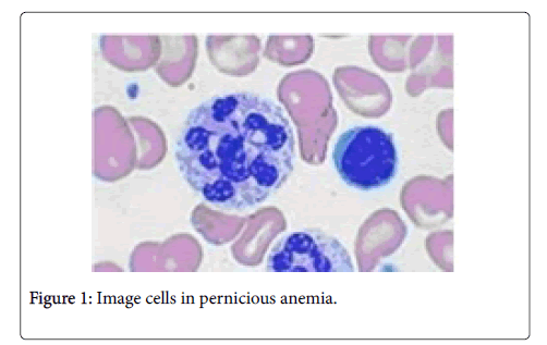
Figure 1 and Figure 2 show two examples of cytological preparations images.
Let's show the work of various methods that allow you to select the outline of objects in the original images. To do this, consider the classical methods-the Prewitt operator and the Roberts operator. These are the most common classical methods for contouring objects in an image. We will also consider the method of wavelet analysis. Then we will compare the results obtained
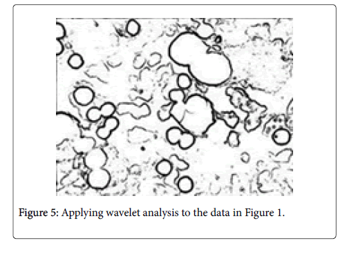
Figure 3 shows the results of using the Prewitt operator, Figure 4 the Roberts operator, Figure 5 the wavelet analysis for the data in Figure 1.
A visual comparison of the data in Figures 3-8 shows that wavelet analysis gives the best results when extracting contours in the original images. At the same time, we used the same algorithms for processing source images (Figures 1 and 2). We see that wavelet analysis has the best unified properties for analyzing different source images. Moreover, we see that wavelet analysis allows us to perform equivalent processing of images that have a uniform background (Figure 1) and a complex background (Figure 2). This is important when processing images of cytological preparations when different staining methods are used.
Wavelet analysis also makes it possible to investigate in detail the structure of the object being analyzed (see in comparison Figures 3-5). This is an important point for research in Clinical and experimental pathology
Conclusion
We considered the possibility and feasibility of using wavelet analysis for image processing of cytological preparations. Shown is a visual comparison between the classical methods of contour detection in the image and the method of wavelet analysis. We have noted the high versatility of the wavelet analysis when selecting the contour of the object in the image. This is important when using different methods for staining cytological preparations. It is also important for the identification of pathology on images of cytological preparations. Thus, the application of wavelet analysis has good prospects for use in research in clinical and experimental pathology.
Disclosure
We declare no conflicts of interest regarding the publication of this paper
Funding
The authors received no financial support for the research, authorship and or publication of this article.
References
- Butler HJ, Ashton L, Bird B, Cinque G (2016) Using Raman spectroscopy to characterize biological materials. Nature protocols 11: 664-687.
- Rowe C, Sitch AJ, Barratt J, Brettell EA (2019) Biological variation of measured and estimated glomerular filtration rate in patients with chronic kidney disease. Kid Int 96: 429-435.
- Sharma AK, Toussaint ND, Elder GJ, Masterson R (2018) Magnetic resonance imaging-based assessment of bone microstructure as a non-invasive alternative to histomorphometry in patients with chronic kidney disease. Bone 114: 14-21.
- Frossing S, Nylander MC, Chabanova E, Kistorp C, Skouby SO (2018) Quantification of visceral adipose tissue in polycystic ovary syndrome: dual-energy X-ray absorptiometry versus magnetic resonance imaging. Acta Radiologica 59: 13-17.
- Abdulhay E, Mohammed MA, Ibrahim DA, Arunkumar N, Venkatraman V (2018) Computer aided solution for automatic segmenting and measurements of blood leucocytes using static microscope images. J Med Sci 42: 58.
- Saieg MA, Troncone G, Zeppa P (2018) Cytological preparations for molecular analysis: a review of technical procedures, advantages and limitations for referring samples for testing. Cyto pathol 29: 125-132.
- Chain K, Legesse T, Heath JE, Staats PN (2019) Digital imageâ€assisted quantitative nuclear analysis improves diagnostic accuracy of thyroid fineâ€needle aspiration cytology. Cancer cytopathol 127: 501-513.
- Dey N, Ashour AS, Ashour AS, Singh A (2015) Digital analysis of microscopic images in medicine. J Adv Microsc 10: 1-13.
- Lyashenko V, Babker MA, Lyubchenko V (2017) Wavelet Analysis of Cytological Preparations Image in Different Color Systems. OALib Journal 4: 3760.
- Babker A, Lyashenko V (2018) Identification of megaloblastic anemia cells through the use of image processing techniques. Int J Clin Biomed Res 4: 1-5.
Citation: Babker MA, LyashenkoV (2020) Wavelet Analysis as Research Tool Image Cytological Preparations. J Clin Exp Pathol 10: 382.
Copyright: © 2020 Babker MA. This is an open-access article distributed under the terms of the Creative Commons Attribution License, which permits unrestricted use, distribution, and reproduction in any medium, provided the original author and source are credited.
Share This Article
Recommended Journals
Open Access Journals
Article Usage
- Total views: 1591
- [From(publication date): 0-2020 - Dec 19, 2024]
- Breakdown by view type
- HTML page views: 1007
- PDF downloads: 584


